


Lung Cancer, Small-Cell (SCLC)
Compliance rules
1Summary
Lung cancer is the third most common malignant tumor in women and the second most common in men in German-speaking countries. In both men and women, lung cancer is the leading cause of cancer-related death. The median age at diagnosis is approximately 70 years. The main risk factor is smoking.
Small cell lung cancer (SCLC) accounts for about 12-15% of all lung cancers. In Germany, approximately 7,000-8,000 people are diagnosed annually. The disease is characterized by a high rate of cell division and rapid progression. These biological characteristics explain the tumor’s high sensitivity to chemotherapy and radiation therapy. On the other hand, they also lead to early dissemination and high recurrence rates. According to the American Cancer Society, the 5-year survival rate for SCLC is 9% (for non-SCLC: 32%) [76]. In stages I-III (Very Limited Disease, Limited Disease), curative treatment is possible. Treatment at these stages is multimodal, incorporating Surgery, systemic tumor therapy, and radiation therapy. For adult patients with Limited Disease whose disease has not progressed following platinum-based chemoradiotherapy, consolidation therapy with durvalumab for 2 years is recommended. The combination of chemotherapy and immunotherapy is the current standard of care in first-line treatment for extensive-stage (ED) SCLC. With combined chemo-immunotherapy, 15-20% of patients achieve a 3-year survival rate.
2Basics
2.1Definition and Basic Information
Lung carcinomas are malignancies arising from the epithelial cells of the respiratory tract. Based on cellular differentiation, a distinction is made between SCLC and NSCLC; among NSCLCs, further differentiation is made according to immunohistological and, more recently, molecular parameters, so that this term should be understood as an umbrella term primarily for adenocarcinomas and squamous cell carcinomas. SCLC, on the other hand, is a distinct entity within the group of neuroendocrine neoplasms (NENs) [57].
The lung is a common site for metastases from numerous malignancies. These, along with other rare pulmonary tumors and benign space-occupying lesions, must be ruled out through the medical history and, if necessary, by histopathological examination.
SCLC develops from neuroendocrine cells in the bronchi. These cells play a regulatory role in the lungs and produce hormones and peptides. Exposure to carcinogens (tobacco components, radon, arsenic, etc.) leads to mutations in tumor suppressor genes and/or proto-oncogenes, which play a central role in aggressive tumor growth [62]. SCLC is characterized by biallelic inactivating mutations in the two tumor suppressor genes TP53 and RB1, which are found in nearly all patients with SCLC [5, 6]. In addition, mutations in the NOTCH gene family or amplifications of the MYC gene are frequently observed in SCLC [3, 56, 61].
Microscopically, SCLC typically presents as a small, blue, round-cell tumor and must be distinguished by conventional histology from other such tumors, such as Ewing sarcoma or lymphomas. The tumor cells are small and have a high nucleus-to-cytoplasm ratio. The cell nuclei appear hyperchromatic with fine-grained chromatin (the so-called “salt and pepper” pattern); a nucleolus is usually barely detectable. Due to the high mitotic activity, apoptosis and pronounced areas of necrosis are frequently observed. The growth pattern is nests, diffuse, or trabecular. To confirm the diagnosis, neuroendocrine markers are typically stained using immunohistochemistry. SCLC tumor cells are generally positive for CD56 (NCAM), synaptophysin, chromogranin A, and TTF-1. Characteristically, a high Ki-67 proliferation rate of over 80% is frequently observed. In contrast to NSCLC, no glandular structures (adenocarcinomas) or keratinization (squamous cell carcinomas) are present. Due to the high proliferation rate, elevated levels of neuron-specific enolase (NSE) and lactate dehydrogenase (LDH) are frequently found in the blood as markers of the high apoptosis rate.
The following statements on epidemiology, risk factors, prevention, and early detection apply to all forms of lung cancer. The subsequent sections of this guideline focus on primary small-cell lung cancer. The first description of small-cell lung cancer is attributed to observations made among workers at the Schneeberg mines in the Ore Mountains [1].
2.2Epidemiology of SCLC in Germany
The following findings are based on data from cancer registries in all federal states, which are regularly compiled at the Center for Cancer Registry Data for nationwide analyses.
During the period 2020-2022, SCLC accounted for approximately 15% of all lung cancer cases in Germany reported to the cancer registries by hospitals, medical practices, or pathology departments; in about 5% of cases, classification was not possible due to nonspecific histological information.
During the same period, approximately 3,500 women and 4,500 men were first diagnosed with SCLC each year. Since the approximately 12% of cases identified in the registry solely through death certificates (DCO) generally do not allow for histological differentiation and are therefore included in the overall lung cancer incidence but not in that of the two subgroups, the figures cited should be understood as minimum estimates.
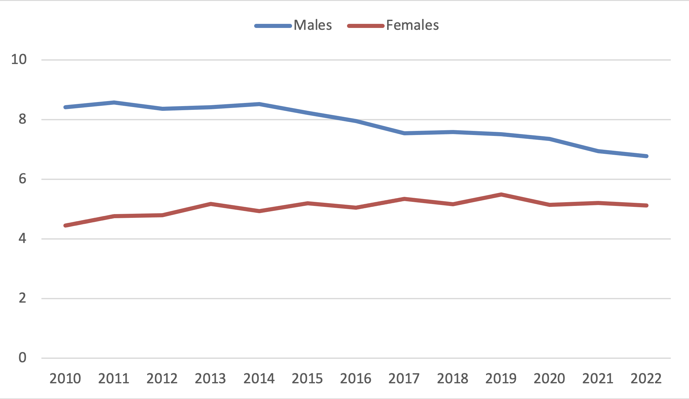
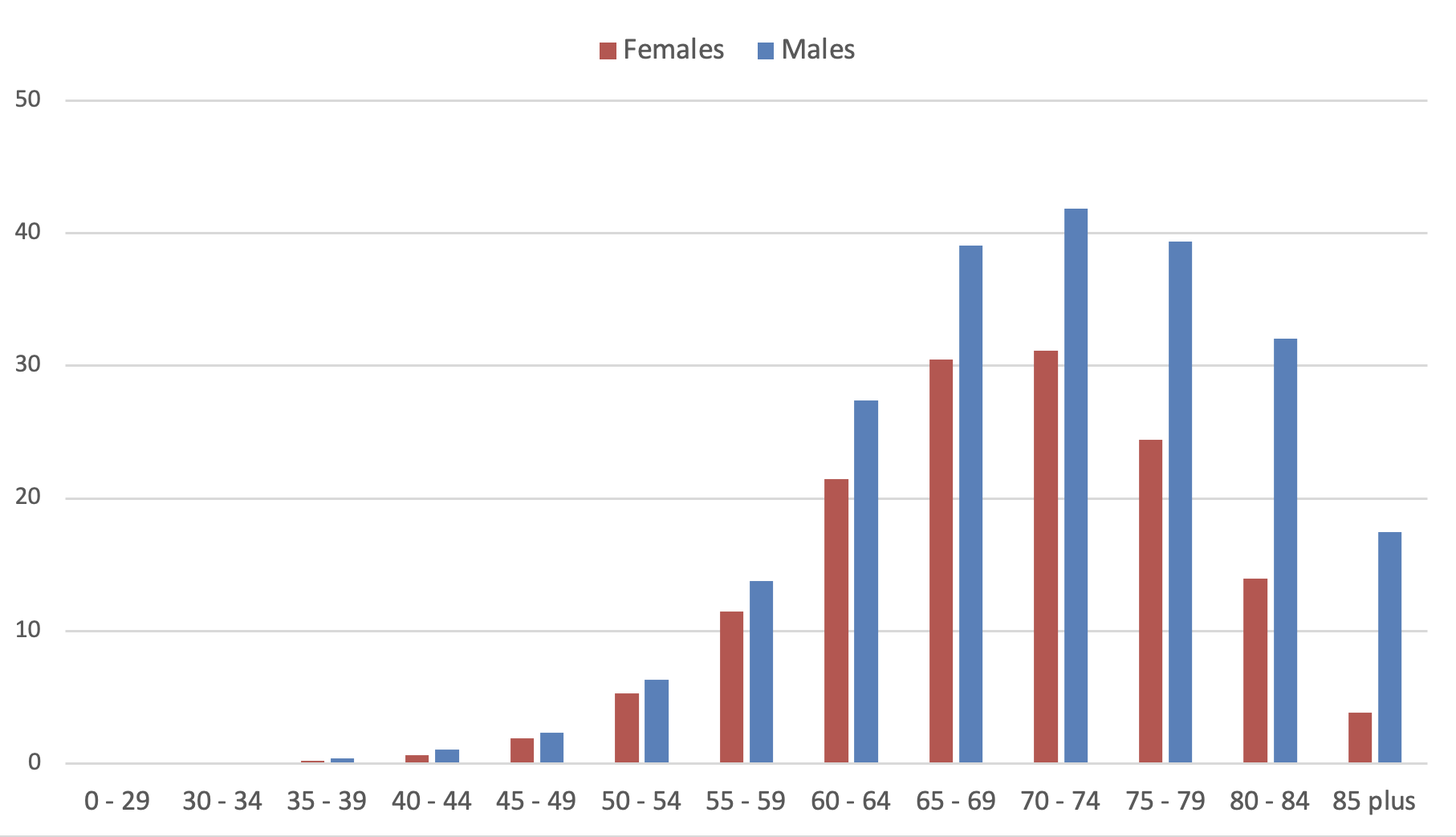
Age-standardized incidence rates are declining among men, while they appear to have plateaued among women (Figure 1). The age-specific incidence increases with age up through the eighth decade of life. The median age was most recently 68 years; only about 2% of those affected are diagnosed before the age of 50 (Figure 2). Incidence rates are declining among men in all age groups as well as among younger women; among women over 60, however, they are still rising. These developments reflect gender-specific trends in smoking behavior with a latency of several decades; in the medium to long term, therefore, a decline is also expected among women. The absolute number of cases has also been declining slightly overall in recent years.
In approximately 75% of new cases (women: 73%, men: 76%) with adequate documentation of tumor staging, distant metastases are already present at the time of the initial SCLC diagnosis. The most commonly affected sites are the liver (44% of cases with distant metastases), the brain (34%), and the bones (27%). On average, two metastatic sites are reported per case. Only just under 5% of cases are diagnosed in the early stages I or II according to the UICC classification.
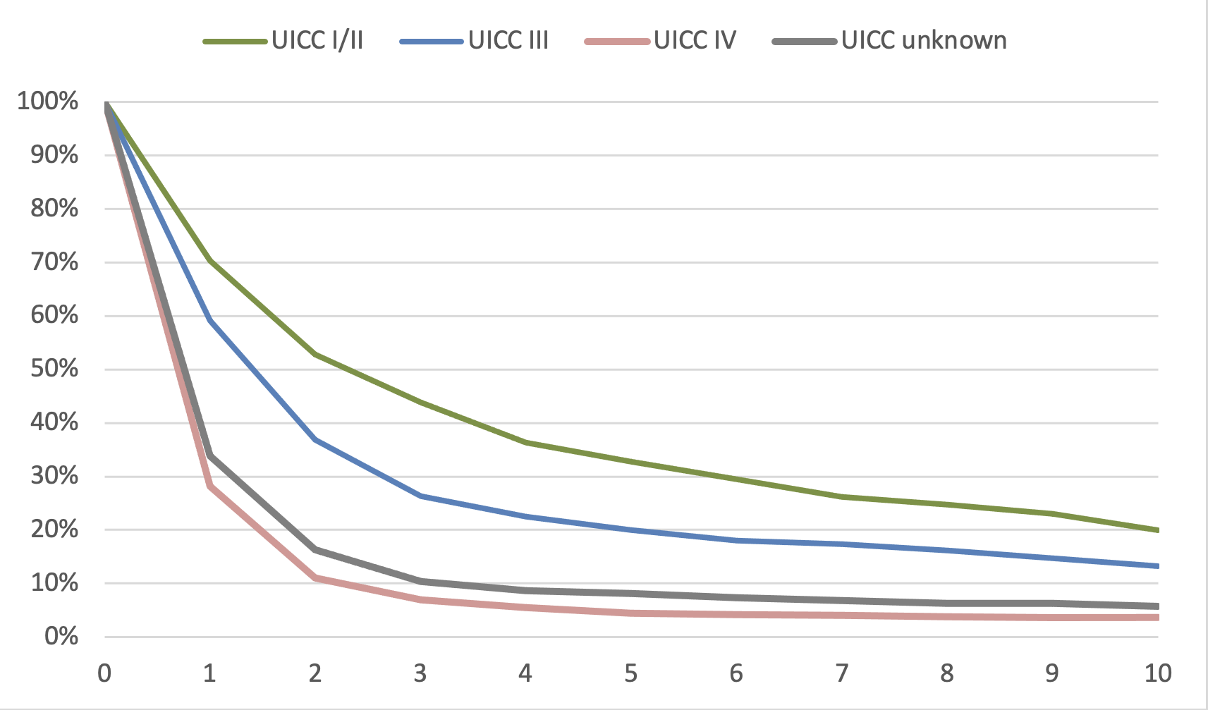
The relative 5-year survival rates - used as an estimate of disease-specific survival - for SCLC in the 2020-2022 period stand at 8.9%, only slightly higher than 10 years earlier (8.5%).
Figure 3 illustrates how survival prognoses depend on tumor stage. The prognosis, which is once again significantly worse compared to NSCLCs, can be explained in part by the less favorable distribution of tumor stages; however, even in the rare cases involving early stages, the outcomes are worse than for NSCLC.
2.3[Chapter not relevant]
2.4Risk Factors
The risk of developing lung cancer is increased by the following factors:
Acquired, exogenous
Smoking, including secondhand smoke
Inhalation of vapors (“vaping”) from electronic cigarettes containing liquids with chemical carcinogens such as acetaldehyde and formaldehyde
Ionizing radiation (high environmental radon exposure, uranium mining, medical radiation exposure)
Particulate matter
Diesel engine exhaust
Asbestos
Quartz dust
Chronic infections
Genetic, endogenous
Individuals with a family history of lung cancer in one or more first-degree relatives have an increased risk of developing the disease.
Further information on risk factors for lung cancer can be found under Non-Small Cell Lung Cancer (NSCLC).
2.5Pathology and Molecular Subgroups
Neuroendocrine tumors (NETs) store active neuropeptides such as bombesin, calcitonin, or serotonin in intracellular granules and release them in response to neural, chemical, or mechanical stimuli. They frequently occur in the gastrointestinal tract, but also in the lungs. Among these, SCLC is the most common type of primary pulmonary neuroendocrine tumor (pNET; [55]). Another, though rarer, high-grade pNET - accounting for approximately 3% of all lung cancers - is large-cell neuroendocrine carcinoma (LCNEC). In addition, there are low-grade carcinoids (approximately 2%, with a ratio of typical to atypical cells of 10:1). The criteria for classifying neuroendocrine tumors of the lung and the gastrointestinal tract do not align.
To diagnose small-cell neuroendocrine lung carcinoma based on biopsies, EBUS, or cytology, immunohistochemical detection of at least two neuroendocrine markers (TTF-1, CD 56, synaptophysin, chromogranin) is required. The Ki67 proliferation rate should exceed 70% Ki67-positive cells. It is particularly important to differentiate this condition from small-cell, basaloid squamous cell carcinomas or non-Hodgkin lymphomas.
Inactivating mutations in the tumor suppressor genes TP53 and RB1 are found in nearly all SCLCs and can be understood here as a fundamental causal mechanism in malignant transformation. Other molecular aberrations found in some cases include mutations in TP73, CREBB genes from the NOTCH family, and, more rarely, in other oncogenes and tumor suppressor genes [3]. The identified molecular aberrations are not yet amenable to targeted therapy.
Based on gene expression analyses in human and murine tumors, differential expression of the four key transcription factors achaete-scute homologue 1 (ASCL1 = ASH1), neurogenic differentiation factor 1 (NeuroD1), and POU class 2 homeobox 3 (POU2F3) was identified, and a new classification was proposed accordingly [4, 65]. To date, this classification cannot be used to guide treatment decisions.
EGFR-mutated NSCLC with secondary SCLC transformation is addressed in the Onkopedia guideline on NSCLC.
3Prevention and Early Detection
3.1Prevention
General recommendations for prevention are based on the risk factors identified to date and personal lifestyle; see Non-Small Cell Lung Cancer (NSCLC):
Avoiding smoking is by far the most important measure
Avoid secondhand smoke and e-cigarettes
Avoid occupational exposure to hazardous substances
Structural measures to reduce radon exposure in high-risk areas
Physical activity
Increased consumption of fruits and vegetables
Avoiding smoking is the most important preventive measure (WHO Framework Convention on Tobacco Control) [6]. Increased consumption of fruits and vegetables reduces the risk of lung cancer, especially among smokers.
3.2Early Detection
There is no recognized early detection method for small-cell lung cancer in Europe in the form of national screening programs; see non-small-cell lung cancer (NSCLC). In Switzerland, the Swiss Accident Insurance Fund (SUVA) offers a screening program to insured individuals with occupational exposure to asbestos, based on the NLST criteria.
4Clinical Characteristics
The clinical symptoms of patients with SCLC do not differ fundamentally from those of patients with NSCLC; see non-small cell lung cancer (NSCLC). Typical features include origin in the central airways and an often short history of tumor-related symptoms such as dyspnea, cough, or signs of upper airway obstruction. A distinctive feature of SCLC is the more frequent occurrence of paraneoplastic syndromes, most commonly involving endocrine manifestations. Table 1 shows the frequency and distribution of paraneoplastic syndromes in patients with lung cancer. The leading symptom of SIADH is hyponatremia; in ACTH syndrome, the characteristic clinical picture of Cushing’s syndrome is often not fully developed due to the clinically short duration of onset. Lambert-Eaton syndrome manifests clinically as muscle weakness accompanied by dysarthria, dysphagia, and proximal limb paresis. Antibody testing (anti-Hu-ANNA-1, anti-neuronal antibody type 1; anti-Ri (ANNA-2, anti-neuronal antibody type 2); anti-CRMP5; anti-Ma1; anti-amphiphysin, among others [7]) can confirm the clinical suspicion of a neurological paraneoplastic syndrome.
Syndrome | SCLC (% of patients) | NSCLC (% of patients) |
|---|---|---|
SIADH | 10 | < 0.1 |
Cushing (ACTH) | 2-4 | < 0.1 |
Lambert-Eaton syndrome | 1 | < 0.1 |
Other neuropathies | up to 5 | < 0.1 |
Clubbing | < 1 | 5 |
Osteoarthropathy | < 1 | 5 |
Hypercalcemia | < 1 | up to 10 |
5Diagnosis
5.1[Chapter not relevant]
5.2Diagnostics
5.2.1Initial Diagnosis
The first step is to confirm the suspected clinical and/or imaging diagnosis; see Figure 4.
Diagnostic workup should continue until metastasis is confirmed or ruled out and, in the absence of metastasis, until the TNM classification is determined; see Table 2.
2 MRI = magnetic resonance imaging;
3 Alternative for peripheral masses: brush, needle, etc.;
4 FDG-PET-CT = 18F-fluorodeoxyglucose positron emission tomography with computed tomography;
5 EBUS = endobronchial or endoesophageal ultrasound with fine-needle biopsy;
6 CM = contrast medium;
7 if there is a high probability of a diagnosis of NSCLC or SCLC
As a result of the FDG-PET-CT examination, a significant percentage of patients are reclassified from stage LD to ED. In 8 studies involving a total of 138 LD SCLC patients, the stage changed to ED in 29 cases, representing an average of 20% of patients [8]. This justifies performing a PET-CT scan prior to planned curative therapy using concurrent chemoradiotherapy or surgery [9, 66].
5.3Classification
5.3.1[Chapter not relevant]
5.3.2Stages
Since January 1, 2025, the criteria of the 9th edition of the TNM classification for lung cancer are effective [53, 54]; see Tables 3 and 4.
For classification, the distinction between “Limited” and “Extensive” disease, developed in 1957 by the Veterans Administration Lung Study, has been used for many decades [11]; see Table 5.
This classification was primarily based on the feasibility of radiation therapy. “Limited Disease” (LD) is defined as tumor spread that can be completely encompassed and irradiated within a tolerable radiation therapy target volume. A further distinction is made by subdividing the LD stage into a “Very Limited Disease” (VLD) group without evidence of mediastinal lymph node involvement and an LD group with mediastinal lymph node involvement.
Although the VA classification is usually sufficient for clinical purposes, the differentiated classification based on the TNM and UICC criteria [10] is now recommended to standardize staging and because of its greater prognostic value. The correspondence between TNM features and the Veterans Administration Lung Study classification is summarized in Table 6.
Stages of the Veterans Administration Lung Study | Correspondence to the TNM Classification |
|---|---|
Very Limited Disease | T1-2 N0-1 |
Limited Disease | T3-4 and/or N2-3 |
Extensive Disease | M1 |
More recently, a new classification has been proposed based on gene expression analyses in human and murine tumors [4]. This classification is based on the differential expression of four key transcription factors: achaete-scute homologue 1 (ASCL1 = ASH1), neurogenic differentiation factor 1 (NeuroD1), and POU class 2 homeobox 3 (POU2F3).
Accordingly, the new classification divides SCLC into the subtypes SCLC-A, SCLC-N, SCLC-P, and SCLC-Y. SCLC-I (inflamed gene signature) has been proposed as an additional subtype [5]. The distinguishability of these subtypes and their therapeutic relevance are the subject of current research and discussion. Initial data suggest greater efficacy of immunotherapy in the “inflamed” subgroup.
5.4[Chapter not relevant]
5.5[Chapter not relevant]
5.6General Condition and Comorbidities
Treatment options for patients with lung cancer are often limited by poor general condition as well as cardiovascular, pulmonary, or other comorbidities, including those related to age. This applies to both curative and palliative therapy. Parameters for assessing operability can be found in non-small cell lung cancer (NSCLC).
To objectively assess general condition in older patients, the use of geriatric assessment tools is recommended; see the Geriatric Assessment Knowledge Base. Tests designed to objectively measure mobility and comorbidities are particularly suitable. The decision to perform additional tests is based on clinical judgment and the planned treatment.
6Treatment
6.1Treatment Structure
6.1.1First-Line Therapy
Treatment recommendations are based on the UICC staging system. However, the conventional classification into Very Limited, Limited, and Extensive Disease (VLD, LD, and ED) is retained when describing treatment options, as treatment studies have generally been conducted based on this classification and it therefore forms the basis for treatment recommendations.
The form of treatment varies depending on the stage. For VLD, the most effective treatment is Surgery combined with curative-intent chemotherapy. In combination with Surgery and/or radiation therapy, curative intent is also achievable for LD through chemoradiotherapy followed by immunotherapy; for ED, in addition to palliative symptom relief, a significant improvement in survival time is now achieved for a portion of patients.
An algorithm for first-line therapy is shown in Figure 5. Whenever possible, patients should be treated as part of clinical trials.
curative intent, palliative intent
PCI = Prophylactic Cranial Irradiation; RT = radiation therapy; hyperfractionated RT = hyperfractionated radiation therapy twice daily, conventional RT = conventionally fractionated radiation therapy once daily, Gy = Gray, CR = complete remission, PR = partial remission, MRI = magnetic resonance imaging, TVD = target volume dose
6.1.1.1Stage I-IIA (Very Limited Disease, VLD)
Only about 5% of patients with SCLC are diagnosed at stages I and IIA (tumors smaller than 5 cm without lymph node involvement). In most cases, these are patients who undergo surgery for a peripheral round lesion, and it is only the histology that reveals the presence of SCLC. In a database analysis of the U.S. National Cancer Database, 1,574 patients were identified and evaluated who received various forms of follow-up treatment after such a resection [12]. Following surgery alone, the 5-year survival rates were 40% (n=388); following additional adjuvant chemotherapy, 52% (n=544); and following additional prophylactic cranial irradiation (PCI), just under 70% (n=99). Mediastinal postoperative radiotherapy (RT) did not provide any additional survival benefit. Based on the retrospective data, adjuvant chemotherapy consisting of 4 cycles of cisplatin/etoposide can be recommended following surgical resection; however, the conclusions regarding PCI are limited due to the small number of cases and possible patient selection bias.
The database analysis by Raman et al. [13] examined the required extent of resection in a total of 1,948 surgically treated SCLC cases at stage T1-2N0. These patients underwent either a wedge resection (n=609), a segmental resection (n=96), or a lobectomy (n=1,233). 75% of the patients were stage IA, 10% were stage IB, and 15% were stage II. 35% of the patients received adjuvant chemotherapy, and 10% received additional PCI. The five-year survival rates were 31% and 35% for wedge resection and segmental resection, respectively; the rate for lobectomy was significantly higher at 45%. Therefore, the primary surgery should be a lobectomy with systematic lymphadenectomy.
If a VLD-stage SCLC is detected via classic diagnostic methods before the initiation of therapy, combined concurrent chemoradiotherapy (CRT) is available as a treatment option in addition to primary surgery with adjuvant therapy.
This treatment modality and its outcomes are discussed in detail in Chapter 6.1.1.2.
Unfortunately, there are no stage-specific randomized comparisons between the two treatment modalities - surgery and concurrent CRT. Two older randomized studies randomized patients who had received neoadjuvant chemotherapy alone to either surgery followed by RT or RT alone. In 146 and 69 randomized patients, respectively, no difference was observed between the arms. In case series and phase II studies, 5-year survival rates of 50-70% were observed for such a neoadjuvant treatment strategy in patients with stage N0, and rates of 35-40% in patients with stage N1.
The value of PCI in stages N0-1 has not been established. However, registry data suggest that PCI increases the 5-year survival rate following surgical resection. Its use must be discussed on a case-by-case basis. The role of adjuvant immunotherapy with durvalumab in the postoperative setting has not been evaluated, but may be considered by analogy with the approach used for LD following CRT.
6.1.1.2Stages IIB and III (Limited Disease, LD)
Approximately one-third of patients with SCLC are classified as having limited-stage disease at first diagnosis (tumors with T3 or T4 features or N1/N2/N3 involvement). These patients are eligible for curative treatment. The 5-year survival rates range from 30-35%. The standard of care is concurrent CRT therapy.
The most effective chemotherapy regimen is the combination of cisplatin and etoposide over 4 cycles. Cisplatin/etoposide can be administered concurrently with RT without dose restrictions and with a tolerable side-effect profile. Cisplatin has a well-documented radiosensitizing effect; fewer data are available for carboplatin, but cohort studies show at least no clear inferiority [67]. The standard dose of cisplatin should be 75-90 mg/m²on day 1, but may also be divided into 25-30 mg/m2 spread over days 1-3. For patients who are not eligible for cisplatin, carboplatin is an alternative. RT should be initiated no later than at the start of the third cycle.
The preferred RT options are hyperfractionated, accelerated radiation therapy at 1.5 Gy twice daily up to a total dose of 45 Gy (up to 60 Gy in phase II studies) or conventionally fractionated, once-daily radiation therapy at 1.8-2.0 Gy per fraction and a total dose of up to 66 Gy. A randomized comparison of these two approaches in the CONVERT study by Faivre-Finn et al. [15] showed no significant improvement with the conventional regimen; the 3-year survival rate in the study was 43% for hyperfractionated RT and 39% for conventional RT. Similarly, the CALGB study by Bogart et al. [16] showed no significant differences. 638 patients received either concurrent CRT with twice-daily RT up to 45 Gy or once-daily RT with a target volume dose (TVD) of 70 Gy. 60% received RT using IMRT. RT was initiated with the first cycle of chemotherapy in 45% of patients; cisplatin was used as the basis of chemotherapy in 81% of cases. Median survival was just under two and a half years, with a five-year survival rate of 29% in the twice-daily RT arm and 34% in the once-daily radiation arm. The rate of side effects did not differ; esophageal complications occurred in 17% of patients.
A dose-escalated hyperfractionated RT regimen with twice-daily hyperfractionated RT up to 60 Gy was used in the randomized phase II study by Grønberg et al. [17]. A total of 176 patients were treated; the two-year survival rates were 74% at 60 Gy compared with 48% at 45 Gy. The most common side effects were hematologic in nature, with neutropenia occurring in 80% of cases. A neutropenic infection was observed in 27%. The esophagitis rate was 21% vs. 18%. In both treatment arms, three patients died from treatment-related complications. This approach is not yet a standard of care; validation in a randomized phase III trial is pending.
Dose-intensified AHF therapy may contribute to improved survival [17, 64]. This opens up the possibility of increasing the effectiveness of treatment for selected patients, e.g., those with bulky tumors.
Table 7 below provides an overview of the results from randomized trials comparing conventional versus hyperfractionated RT.
Authors | n | Treatment | 3-Year OS | 5-Year OS |
|---|---|---|---|---|
Turrisi [14] | 206 | RT 45 Gy, 1.8 Gy per fraction x 25 | 33% | 16% |
211 | RT 45 Gy, 2 × 1.5 Gy ED × 15 | 27% | 26% | |
Faivre-Finn CONVERT [15] | 270 | RT 66 Gy, 1 × 2 Gy ED × 33 | 39% | 27% |
273 | RT 45 Gy, 2 × 1.5 Gy ED × 15 | 43% | 33% | |
Bogart | 325 | RT 70 Gy, 1 x 2 Gy dose per fraction x 35 | 44% | 34% |
313 | RT 45 Gy, 2 × 1.5 Gy per fraction × 15 | 42% | 29% | |
Grønberg [17] | 89 | RT 60 Gy, 2 × 1.5 Gy per fraction × 20 | 74% (2 years) | 42% (4 years) |
81 | RT 45 Gy, 2 × 1.5 Gy per fraction × 15 | 48% (2 years) | 28% (4 years) | |
Yu TRISS [64] | 108 | RT 54 Gy, 2 × 1.8 Gy per fraction × 15 | 76% (2 years) | NA |
116 | RT 45 Gy, 2 × 1.5 Gy per fraction × 15 | 54% (2 years) | NA |
6.1.1.2.1Maintenance Immunotherapy Following Concurrent CRT
The results of the ADRIATIC trial in SCLC stages I-III showed that durvalumab, as consolidation therapy following cCRT and in the absence of disease progression, led to a statistically significant and clinically meaningful improvement in both PFS and OS compared with placebo. The median OS was 55.9 months in the durvalumab arm compared with 33.4 months in the placebo arm (hazard ratio [HR] 0.73; 95% confidence interval [CI]: 0.57-0.93; p = 0.0104). The median PFS was 16.6 months for durvalumab and 9.2 months for placebo (HR 0.76; 95% CI: 0.61-0.95). The safety analysis showed that the rate of pneumonitis was slightly increased with durvalumab (38% vs. 30% in the placebo arm). The ADRIATIC study thus demonstrates that consolidation therapy with durvalumab for 2 years following successful cCRT in patients with LD-SCLC leads to a significant prolongation of survival and exhibits an acceptable safety profile [54]. Therefore, the ADRIATIC regimen may be considered the standard of care for stage I-III LD-SCLC following chemotherapy and RT. Approval has been granted by the EMA for both combined and sequential CRT. In patients treated with RT according to the Turrisi protocol, durvalumab did not result in a clear survival benefit.
6.1.1.2.2Explanatory Notes on Concurrent CRT
Simultaneous CRT is superior to the sequential approach and is therefore the preferred treatment option, provided it can be administered in a timely manner and at the planned dosage [68].
A sequential approach should therefore only be used as part of individualized treatment plans in the presence of contraindications to concurrent CRT.
Protocols containing carboplatin have not been sufficiently tested in an adjuvant setting or as part of concurrent CRT regimens and should therefore be used here only in cases of clear contraindications to cisplatin. Initial chemotherapy with carboplatin and etoposide followed by consolidating RT may be a treatment option for patients in significantly compromised general condition if standard therapy with cisplatin and etoposide is not feasible.
Another possible treatment option is to perform concurrent hyperfractionated CRT with cisplatin/etoposide in the first cycle and concurrent RT at 2 × 1.5 Gy per day starting on the first day of treatment up to a total dose of 45 Gy, followed by a switch to the cisplatin/irinotecan combination for the remaining three cycles of chemotherapy alone. In a Japanese patient cohort, this approach was found to be equivalent to the standard approach involving continued cisplatin/etoposide [19].
The use of anthracycline-containing regimens should be avoided in the context of concurrent CRT due to their lower efficacy and higher toxicity. Similarly, dose-intensification strategies are not recommended outside of clinical trials.
6.1.1.3Prophylactic Cranial Irradiation (PCI) in Stage LD
PCI reduces the risk of brain metastases from 40% in patients who do not receive PCI to less than 10% in patients who do, and improves the 5-year survival rate by 5% in absolute terms [20].
PCI is therefore an established component of therapy for patients following concurrent CRT.
PCI may be associated with impairments in cognitive function. Several studies have therefore sought to reduce this side effect by sparing the hippocampus. A Spanish study by Rodríguez de Dios et al. [21] included 150 patients; 75 received classic PCI with 25 Gy in 10 fractions, and the other half received the same PCI with hippocampal sparing. This study demonstrated better preservation of neurocognitive function with hippocampal sparing. The rates of significant deterioration were 8.7% vs. 20.6%. A second study from the Netherlands by Belderbos et al. [22] included 168 patients. Here, too, 25 Gy in 10 fractions was administered with and without hippocampal sparing. The rates of significant deterioration in neurocognitive function were 29% vs. 28%, respectively, and thus did not differ. In both studies, the rate of new brain metastases did not differ, and survival rates were also the same.
Hippocampal sparing therefore does not reduce the efficacy of PCI and does not impair survival. However, its effect on the preservation of neurocognitive function has not been conclusively established [49].
6.1.1.4Extensive Disease (ED)
60-70% of patients with SCLC are at the extensive disease stage at first diagnosis. The standard of care is systemic tumor therapy with chemotherapy and immunotherapy. In addition to improving symptom control and thereby enhancing quality of life, this approach leads to prolonged survival compared with chemotherapy alone. With chemo-immunotherapy, the median survival time for ED patients is approximately 12 months, the 2-year survival rate is 20-25%, and the 3-year survival rate is 15-20%. The addition of immunotherapy has thus tripled the 3-year survival rates of patients compared to chemotherapy alone.
6.1.1.4.1Systemic Tumor Therapy (ED-SCLC)
An algorithm for selecting chemo-immunotherapy in stage IV is shown in Figure 6.
1 ECOG PS = classification of general condition
The results of systemic therapy for extensive-stage disease can be summarized as follows:
Platinum-based treatment regimens achieve significantly higher complete remission rates than non-platinum-based combination therapies. In meta-analyses, the results regarding overall survival (OS) are inconsistent. In a meta-analysis of 5,530 patients, no significant difference was found in OS rates at 6, 12, or 24 months [23].
Regarding the choice of platinum agent, the majority of studies show that cisplatin is slightly more effective than carboplatin. In a relatively small meta-analysis based on individual patient data with heterogeneous treatment regimens, cisplatin and carboplatin were equally effective, and remission rates were the same. Carboplatin has a more favorable side effect profile. The two platinum derivatives are equally effective in the treatment of stage ED.
Achieving the full target dose of platinum is an important prognostic factor.
Protocols containing anthracyclines, such as ACO, EpiCO, or ACE (doxorubicin/adriamycin or epirubicin plus cyclophosphamide plus vincristine or etoposide), are generally effective but are no longer used in primary therapy today due to anthracycline-associated cardiotoxicity, which may be exacerbated by additional radiation therapy.
Dose intensification increases remission rates but does not prolong OS.
Alternating between different combination therapies also does not improve OS compared with sequential therapy.
Several randomized phase III studies are now available comparing chemotherapy alone versus chemotherapy plus PD-(L)1 antibodies.
The IMpower 133 study [25] randomized 403 patients to either 4 cycles of carboplatin/etoposide alone or the same regimen plus the PD-L1 antibody atezolizumab, followed by maintenance therapy with atezolizumab in responders. The remission rates did not differ (60% vs. 64%), but the 12-month PFS rate was significantly higher in the atezolizumab arm at 12.6% vs. 5.4%. Median OS was significantly prolonged by 2 months, from 10.3 to 12.3 months (HR 0.76). The 2-year OS rates were 22% vs. 18%. Longer-term follow-up data are not available.
In the CASPIAN study [26], the anti-PD-L1 antibody durvalumab, in combination with 4 cycles of platinum/etoposide followed by maintenance therapy with durvalumab in responders, also led to an extension of overall survival from 10.3 to 13.0 months compared with 4-6 cycles of platinum/etoposide (HR 0.73). The 2-year OS rates were 22% vs. 14%, and the 3-year OS rates were 18% vs. 6%.
The addition of tremelimumab, a CTLA-4 antibody, to platinum/etoposide and durvalumab did not improve patient OS in the CASPIAN trial.
The Keynote 604 trial [27] evaluated pembrolizumab as an add-on to platinum and etoposide. Although this trial was not statistically significant in terms of OS (HR 0.80, p=0.016), the 2-year survival rates were 23% vs. 11% in this trial as well.
The ASTRUM study [28, 70] evaluated the PD-1 antibody serplulimab in combination with platinum and etoposide. A total of 585 patients, predominantly from Asia, were enrolled in this study. Median OS was significantly prolonged at 15.8 vs. 11.1 months, and remission rates were also higher with immunotherapy. The 4-year OS rates were 21.9% and 7.2%, respectively.
The CAPSTONE-1 phase III trial [29], which was also conducted exclusively in China, demonstrated an OS benefit for the anti-PD-L1 antibody adebrelimab in combination with carboplatin and etoposide compared to chemotherapy alone (15.3 months vs. 12.8 months; HR 0.72). To date, no approval has been granted for Europe (as of August 2026).
The RATIONALE-312 phase III trial [69], which was also conducted exclusively in China, demonstrated an OS benefit for the anti-PD-L1 antibody tislelizumab in combination with etoposide and cisplatin or carboplatin compared with chemotherapy alone (15.5 months vs. 13.5 months, HR 0.75).
All studies incorporating PD-(L)-1 inhibitors thus demonstrate a benefit for immunotherapy, making the combination of approved PD-(L)-1 inhibitors the current standard of care in first-line therapy.
Atezolizumab is approved for first-line therapy in combination with carboplatin and etoposide, and durvalumab is approved in combination with cisplatin or carboplatin plus etoposide. In the CASPIAN trial, the addition of durvalumab to cisplatin/etoposide was 10% more effective compared to carboplatin/etoposide. It is not clear from this setting whether selection effects or drug interactions contribute to this difference. Serplulimab is approved in combination with carboplatin and etoposide; tislelizumab is approved in combination with etoposide and platinum-based chemotherapy.
According to the approval, there is no limit on the duration of therapy; the combination of chemotherapy and immunotherapy should be administered over 4 (up to a maximum of 6) cycles, after which immunotherapy is continued until disease progression.
In patients with humoral-mediated paraneoplasias (Lambert-Eaton syndrome, other neuropathies), the indication for immune checkpoint inhibitor therapy should be viewed with caution; if necessary, treatment should not begin until the paraneoplasia has resolved.
The study results for primary combined chemo-immunotherapy are shown in Table 8 below
Study | Arm | n | RR | PFS (mo) | HR PFS | OS (mo) | OS 24 mo | OS 36 Mo | HR OS |
|---|---|---|---|---|---|---|---|---|---|
IMpower-133 [25] | Atezolizumab | 201 | 60 | 5.2 | 0.72 | 12.3 | 22% | 0.76 | |
Placebo | 201 | 64 | 4.3 | 0.62 - 0.96 | 10.3 | 18% | 0.6–0.95 | ||
CASPIAN [26] | Durvalumab | 268 | 68 | 5.1 | 0.80 | 12.9 | 22% | 18% | 0.75 |
Placebo | 269 | 58 | 5.4 | 0.70 - 1.01 | 10.6 | 14% | 6% | 0.68–1.00 | |
KEYNOTE-604 [27] | Pembrolizumab | 228 | 71 | 4.5 | 0.75 | 10.8 | 23% | 0.80 n.s. | |
Placebo | 225 | 62 | 4.3 | 0.61 - 0.91 | 9.7 | 11% | 0.64 - 0.98 | ||
ASTRUM -005 [28.70] | Serplulimab | 389 | 80 | 5.7 | 0.46 | 15.8 | 31.7% | 0.63 | |
Placebo | 196 | 70 | 4.3 | 0.38 - 0.59 | 11.1 | 18.7% | 0.49 - 0.82 | ||
CAPSTONE-1 [29] | Adebrelimab | 230 | 70.4 | 5.8 | 0.67 | 15.3 | 31.3% | 0.72 | |
Placebo | 232 | 65.9 | 5.6 | 0.54 - 0.83 | 12.8 | 17.2% | 0.58 - 0.9 | ||
RATIONALE-312 [69] | Tislelizumab | 227 | 68.3 | 4.7 | 0.65 | 15.5 | 33.2% | 0.78 | |
Placebo | 230 | 61.7 | 4.3 | 0.53 - 0.80 | 13.5 | 22.4% | 0.63 - 0.95 |
The results of the phase III IMforte study were recently published [75]. The study demonstrated a significant prolongation of PFS and OS with lurbinectedin plus atezolizumab compared to atezolizumab alone. Lurbinectedin in combination with atezolizumab can be considered a new option for maintenance therapy in patients with ECOG status 0 or 1 and without brain metastases and has since been approved in the EU for this indication. The potential survival benefit must be weighed against the higher rate of adverse effects.
Currently, the BEAT-SC study is investigating the combination of bevacizumab (angiogenesis inhibitor) with atezolizumab + carboplatin or cisplatin + etoposide (ACE) compared to placebo + ACE every 3 weeks for 4 cycles, followed by maintenance therapy with bevacizumab + atezolizumab every 3 weeks versus placebo + atezolizumab. The addition of bevacizumab to ACE was generally well tolerated, and the safety profile was consistent with the known risks of the individual drugs and the underlying disease. The primary endpoint, PFS, was met and showed a statistically significant improvement in PFS in favor of bevacizumab + ACE compared with placebo + ACE. OS data were immature at the first interim analysis and showed no improvement in favor of bevacizumab + ACE. OS follow-up is ongoing [51].
The efficacy of systemic chemotherapy is lower intracerebrally than outside the CNS. In older studies, chemotherapy alone was associated with shorter survival compared to chemotherapy combined with radiation therapy.
As a rule, therefore, the detection of intracerebral metastases indicates the need for additional RT. The extent and timing of this additional local therapy have become a topic of debate due to recent study results. The FIRE study [30] is a case series of 710 patients with brain metastases from SCLC who were treated with stereotactic RT. Approximately one-third of the patients had 1 brain metastasis, one-third had 2-4, and the remaining one-third had more than 4. The median OS times in the respective groups were 11 months, 8.7 months, and 8.0 months. New brain metastases developed in 55% of patients who initially had a single metastasis and in 70% of patients with multiple brain metastases. A matched-pair analysis comparing patients who received stereotactic RT with those who received whole-brain RT (187 vs. 178 patients) showed a survival advantage for patients treated with stereotactic RT, although the intracerebral recurrence rate - at approximately 60% - was twice as high as that following whole-brain RT (30%). Compared with stereotactic RT plus whole-brain RT, stereotactic RT alone is associated with significantly less impairment of neurocognitive function in patients with 1-3 brain metastases of various etiologies (60% lung carcinomas) [31]. Whole-brain RT can also be administered in the form of hippocampus-sparing RT in patients without metastases in the hippocampal region. The NRG study [32] demonstrated better preservation of neurocognitive function in over 500 patients with brain metastases of various etiologies (60% lung carcinomas), while maintaining equivalent efficacy and survival rates.
In the IMpower 133 study, patients with brain metastases did not benefit from atezolizumab treatment; in the KEYNOTE-604 study, patients with brain metastases actually tended to fare worse in the pembrolizumab group. In CASPIAN, progression-free survival (PFS) is absolutely identical in patients with and without brain metastases; median overall survival is better in the durvalumab group (8.7 vs. 11.8 months), but the curves converge again over time.
While IMpower 133 and KEYNOTE-604 included previously treated (usually radiation-treated) patients with stable brain metastases, 90% of patients with brain metastases in CASPIAN were treatment-naive.
Neither atezolizumab nor durvalumab reduced the incidence of new brain metastases. Approximately 15% of patients without initial brain metastases developed new brain metastases during the course of therapy.
In CASPIAN, among patients with brain metastases, 3 patients in the durvalumab arm and 4 patients in the durvalumab + tremelimumab arm achieved 3-year survival, whereas none of the patients receiving chemotherapy alone achieved this [32].
Therefore, combined chemotherapy and immunotherapy - with the initial omission of additional RT - is an option for asymptomatic patients, as is stereotactic RT for patients with a limited number of brain metastases. Symptomatic patients with multiple intracerebral lesions, however, should continue to receive early whole-brain RT.
In older patients in good general health, the results are comparable to those in younger patients. Age per se is therefore not a negative prognostic factor. To date, there is no evidence that immunotherapy is less effective in older patients. It should be noted that the therapy is associated with higher hematologic toxicity in older patients, which requires dose adjustments.
Only patients with a performance status (PS) of 0 or 1 were included in the studies on combined chemo-immunotherapy. It is unclear whether PS 2 patients benefit from the addition of immunotherapy. Further studies are needed in this regard. The approval does not exclude PS 2 patients. In cases where a PS 2 status is due to tumor burden, the administration of additional immunotherapy is justified despite the lack of study data.
For patients in poor general condition due to significant comorbidities, purely symptom-oriented therapy or, at most, monotherapy with a chemotherapeutic agent is recommended. Monotherapy with an immunotherapy agent has not been tested and should not be used.
Predictors of immunotherapy efficacy have not yet been sufficiently defined. Tumor cells in SCLC rarely express PD-L1; immune cells in the peripheral regions of the tumor are more frequently PD-L1-positive. PD-L1 expression was not predictive of the efficacy of PD-L1 antibodies in either IMpower 133 or CASPIAN; in IMpower 133, PD-L1-negative patients actually tended to benefit more from atezolizumab. In CASPIAN, PD-L1 positivity and the HLA marker DQB1*03:01 were favorable factors for achieving 3-year survival with durvalumab plus tremelimumab. For durvalumab monotherapy, the HLA marker DQB1*03:01 was not predictive. Tumor mutation burden was also not a predictive factor for the efficacy of the PD-L1 antibody in either IMpower 133 or CASPIAN.
Following combined chemo-immunotherapy, immunotherapy should be continued as maintenance therapy, in accordance with the approval, for at least 2 years or until disease progression.
Response to chemo- and immunotherapy can be assessed after at least 2 treatment cycles, typically after 3 to 4 cycles. If there is a response, the combination therapy should be administered for a total of 4 cycles. If tolerability is good and further clinical benefit is expected, an extension to up to 6 cycles is also possible, followed by maintenance immunotherapy.
If there is no response to first-line therapy, the prognosis is very poor. A switch to second-line therapy may be made at an early stage. The DeLLphi-304 study [63] compared tarlatamab, a bispecific T-cell engager, with chemotherapy in patients with SCLC who had progressed during or after initial platinum-based chemotherapy. Tarlatamab resulted in longer survival than chemotherapy (13.6 months vs. 8.3 months, HR 0.6). Tarlatamab has been approved for this indication in the EU since May 2026 and in Switzerland since July 2026.
An important negative prognostic factor is elevated LDH.
Tumor lysis syndrome may occur or be exacerbated at the start of chemotherapy.
6.1.1.5Local Therapeutic Measures in Stage IV (ED-SCLC)
In a randomized EORTC study [35], thoracic adjuvant RT in patients who had not progressed after first-line therapy but had not received primary chemo-immunotherapy did not result in a significant improvement in the primary endpoint of OS (HR 0.84; p=0.066), but it did result in an increase in the 2-year survival rate from 3% to 13%. Female patients under the age of 70 with residual thoracic tumor benefited particularly from the subsequent RT.
Consolidation RT to the primary tumor has not been evaluated when combined chemotherapy and immunotherapy are used as first-line treatment. This was not included in either the IMPOWER-133 or CASPIAN trials. It is unclear whether consolidative RT increases the long-term survival rate in patients with residual thoracic tumors and very good remission of distant metastases, even when combined chemo-immunotherapy is used as first-line treatment. Given the expected thoracic and pulmonary toxicity associated with ongoing immune maintenance therapy, this approach is not a standard procedure and is being evaluated in clinical trials (e.g., Maverick, NCT04155034).
There are varying study findings regarding PCI in patients with extensive disease. In the EORTC study [36] involving patients who showed no progression after first-line therapy and had no clinical signs of brain metastases, PCI resulted in improved OS compared with observation (HR 0.68; median 1.3 months). However, this study did not include systematic cranial MRI follow-ups, and cranial RT in the control arm was initiated only upon the onset of clinical symptoms of CNS involvement. Only 45% of patients in the non-PCI arm received second-line chemotherapy, compared with 68% in the PCI arm; data on the frequency of cranial RT in the control arm are lacking.
A randomized Japanese study [37] included only patients without MRI-confirmed brain metastases. In this study, cranial MRI scans were performed every 3 months in the control arm, and cranial RT was initiated upon imaging evidence of brain metastases. In this study, 89% of non-PCI patients received second-line chemotherapy, and of the 51 patients with newly diagnosed brain metastases, 81% were treated with RT or surgery. In this study, a slight, statistically nonsignificant survival disadvantage was observed with PCI, with a median survival of 11.6 vs. 13.7 months (HR 1.27; p = 0.094).
In selected patients, PCI may be offered as an alternative to monitoring with follow-up MRI scans following a response to systemic therapy and should be discussed on an individual basis.
6.1.2Second-Line Therapy
The indication and selection of second-line therapy depend on the stage of the disease, the patient’s general condition and comorbidities, prior therapy, and the timing of disease recurrence or the duration of the treatment-free period. The algorithm is shown in Figure 7 and Figure 8, distinguishing between local progression (Figure 7) and systemic progression (Figure 8).
Particularly for cases of local recurrence, only retrospective analyses, case series, and clinical experience are available. The recommendations are therefore not supported by prospective studies but reflect a clinically feasible approach.
1 Thoracic radiotherapy while undergoing ongoing immunotherapy has not yet been sufficiently evaluated in clinical trials. A potentially increased risk of pulmonary toxicity should be considered.
1 Treatment regimens: ACO = doxorubicin/epirubicin, cyclophosphamide, vincristine
2 In these situations, tarlatamab is the preferred recommendation, as the other options are less expensive but inferior in terms of OS, PFS, response rate, and quality of life
3 Patients with PS 2 were not included in the studies relevant to approval
4 The recommendation regarding the choice between surgical resection or stereotactic treatment of an isolated adrenal or brain metastasis is based on case reports and clinical experience. It is not supported by prospective studies or case series involving a large number of patients.
In Switzerland, lurbinectedin is approved as a second-line treatment alongside topotecan, provided there are no brain metastases and at least 30 days have elapsed since platinum-based therapy.
6.1.2.1Local and Regional Progression - Second-Line Therapy
If a patient develops an intrapulmonary secondary tumor following surgical resection and adjuvant chemotherapy, the possibility of a secondary tumor with a different histology must also be considered. In the case of a new round lesion, with lymph node involvement or distant metastasis ruled out via PET-CT and, if necessary, further mediastinal staging, a repeat primary resection may be performed. If SCLC is histologically confirmed, it is unclear whether repeat adjuvant chemotherapy offers any benefits.
If primary resection is not performed, histological confirmation should be sought prior to treatment. If SCLC histology is confirmed prior to treatment, concurrent CRT may be performed as an alternative to surgery. For other histologies, histology- and stage-specific therapy should be initiated.
If patients develop a locoregional relapse with mediastinal lymph node involvement following surgical resection and adjuvant chemotherapy, simultaneous chemoradiotherapy may be considered - similar to the approach for LD-SCLC - after histological confirmation and exclusion of distant metastasis via PET-CT.
If, in stage LD, complete remission of lymph node involvement is achieved after completion of concurrent CRT, but the primary tumor persists or shows local progression again, surgical resection of the primary tumor may be considered on a case-by-case basis. Prior to this, N2 or N3 involvement should be ruled out using PET-CT and, if required, further mediastinal staging; cerebral metastasis should also be ruled out using cranial MRI. Pneumonectomy should be avoided. Stereotactic RT may also be considered on a case-by-case basis.
If, following completion of concurrent CRT, a locoregional relapse with mediastinal lymph node involvement occurs, systemic therapy with chemotherapy and immunotherapy - analogous to first-line therapy in stage ED-SCLC - is indicated.
If local progression is observed in a patient with primarily metastatic disease while distant metastases remain stable, local RT of the progressive tumor may be performed. In this case, chemo-immunotherapy or immunotherapy may be continued initially, and a switch to second-line chemotherapy should only occur upon renewed systemic progression. It should be noted that radiation therapy to the primary tumor while undergoing ongoing immunotherapy has not yet been investigated in large-scale studies, and there may be a higher risk of pulmonary toxicity.
6.1.2.2Systemic Progression - Second-Line Therapy
If a solitary adrenal or brain metastasis occurs as a relapse at the initial VLD or LD stage, local therapy is an option. In the case of an adrenal metastasis, this is preferably performed as surgical resection, and in the case of brain metastases, preferably as stereotactic RT. It is unclear whether subsequent systemic chemotherapy improves the prognosis. Given the current metastatic disease status and the positive data on chemo-immunotherapy, additional chemo-immunotherapy, analogous to primary therapy in stage IV, is recommended.
As an alternative to a local approach followed by systemic chemo-immunotherapy, the latter may also be used as the primary treatment. No prospective studies on the value of the local approach are available; the treatment recommendation is based on case reports and clinical experience.
In cases of disseminated progression or relapse, systemic second-line therapy is indicated for patients with an ECOG PS of 0–2 and for those with an ECOG PS of 3 due to the disease. It leads to symptom relief and prolongation of survival. Depending on the timing of a recurrence, the progression may be classified as chemotherapy-sensitive or chemotherapy-refractory. While data from the period prior to the use of PD-L1 inhibitors in first-line therapy suggest chemotherapy-sensitive progression after a 60-day platinum-free interval (the period between the last administration of platinum-based chemotherapy and detected progression), retrospective data from Japan following the introduction of PD-L1 inhibition show a shift in the timing to a 75-day platinum-free interval.
The later progression or relapse occurs, the more effective second-line chemotherapy is and the longer the survival benefit that can be achieved.
The results regarding systemic therapy for extensive disease can be summarized as follows:
6.1.2.2.1Chemotherapy
Drugs with proven efficacy in the second-line setting include topotecan, irinotecan (including the liposomal formulation), paclitaxel, ifosfamide, anthracyclines (including amrubicin), and lurbinectedin.
Topotecan has been compared in a randomized trial against best supportive care [38]. Topotecan resulted in a significant prolongation of survival from 14 to 26 weeks. The benefit was observed in both sensitive and refractory relapses. Oral and intravenous administration of topotecan are equivalent.
Topotecan is currently the only therapy specifically approved for second-line treatment of SCLC and was therefore used as the standard in the comparator arm of studies.
In a study comparing cisplatin/etoposide/irinotecan versus topotecan in patients with sensitive relapse [39], the combination therapy prolonged median OS from 12 to 18 months; however, it did not increase or achieve long-term survival beyond 3 years and was associated with significantly higher toxicity.
Similarly, in the French study by Baize et al. [40], a benefit was observed for re-treatment with carboplatin/etoposide compared to topotecan in patients with sensitive relapse and more than 90 days of treatment-free survival.
Further studies showed no superiority of the ACO protocol or of individual agents such as amrubicin and liposomal irinotecan compared with topotecan therapy.
Similarly, the Atlantis study [41], which compared adriamycin plus lurbinectedin versus topotecan or ACO in the control arm in 600 patients, showed no benefit for the combination. The treatment-free interval had to be at least 30 days. Median OS was 8.6 months in the lurbinectedin arm and 7.6 months in the control arm. The comparison of the survival curves was virtually identical.
Similarly, the RESILIENT study, which compared liposomal irinotecan with topotecan in 461 patients, showed no benefit for the liposomally encapsulated drug [42].
6.1.2.2.2Immunotherapy
Although the administration of immunotherapy with a PD-L1 checkpoint inhibitor as second-line treatment achieved remission rates of approximately 12% in phase II studies (Checkmate 032) [43], it was not successful in randomized trials. In the Checkmate 331 study, there was no difference in PFS or OS between nivolumab and topotecan in the overall population [44].
In the phase III DeLLphi-304 trial, tarlatamab, a bispecific antibody (BiTE molecule) targeting CD3 and DLL3, demonstrated a significant prolongation of PFS and OS compared to second-line chemotherapy (OS 13.6 months vs. 8.3 months, HR 0.6) with an ORR of 35%. There was also a marked reduction in serious adverse effects with tarlatamab compared to standard chemotherapy. The most common adverse events were cytokine release syndrome (CRS), fatigue, and fever, although the frequency of CRS decreased significantly after the first dose. Adverse effects led to treatment discontinuation in 5% of patients (compared to 12% for chemotherapy). The available data suggest that tarlatamab is effective within the brain [71].
Based on phase II data alone, the FDA granted accelerated approval in May 2024 for tarlatamab to treat adults with ED-SCLC, regardless of DLL3 expression, whose disease has progressed following platinum-based chemotherapy and PD-L1 inhibition [48]. Approval in the EU followed in May 2026. Phase III trials are currently evaluating the combination of tarlatamab with durvalumab as first-line maintenance therapy following platinum-containing chemoimmunotherapy for ED-SCLC (NCT06211036, DeLLphi 305) as well as the use of tarlatamab as first-line maintenance therapy following cCRT in LD-SCLC (NCT06117774, DeLLphi 306). Studies on the methodology of tarlatamab administration in an outpatient setting have been published by participating centers [78].
Based on the results of the DeLLphi-301 study [48], tarlatamab appears to be the preferred option for inoperable relapses following chemoimmunotherapy.
7Targeted Agents
Following negative trial results for aflibercept, bevacizumab, thalidomide, vandetanib, and others, antiangiogenic agents are not indicated in either first- or second-line treatment.
Further negative randomized phase II studies are available for mTOR inhibitors, HDAC inhibitors, BCL-2 antisense agents, and PARP inhibitors.
The maintenance therapy trial with the PARP inhibitor niraparib showed no difference between the two treatment arms in either PFS or OS.
7.1Therapeutic Approaches Under Development
7.1.1Innovative immunotherapies
Obrixtamig [73], another bispecific antibody targeting DLL3 and CD3, demonstrated a preliminary ORR of 17% and a DCR of 43% in a phase I trial (NCT04429087); while another study combining obrixtamig with topotecan (NCT05990738) showed a preliminary ORR of 70% and a DCR of 87%.
Ivonescimab, a bispecific antibody against PD-1 and VEGF, demonstrated an ORR of 80% across all dose levels in a first-line phase I study in combination with carboplatin and etoposide, with an acceptable adverse event profile [74]. Bispecific antibodies against PD-1 and VEGF are currently being evaluated in randomized phase III trials (NCT06712355).
Other innovative immunotherapies are currently in early clinical phases, including trispecific antibodies against DLL3 and CD3 (NCT06440057) and CAR-T cells against DLL3 (NCT03392064, NCT05680922).
7.1.2Antibody-Drug Conjugates (ADCs)
Ifinatamab Deruxtecan (I-DXd, DS-7300) is a novel ADC that targets the B7-H3 protein. B7-H3 is a transmembrane protein that is overexpressed in various solid tumors, including SCLC, and is associated with a poor prognosis [72]. In preclinical studies, DS-7300 demonstrated specific binding to B7-H3 and inhibited the growth of B7-H3-expressing cancer cells in vitro. In vivo, it exhibited significant antitumor activity in various xenograft models, including patient-derived xenograft (PDX) models of SCLC [72]. DS-7300 was evaluated in a Phase I/II study (NCT04145622) involving patients with advanced solid tumors, including SCLC. With a median follow-up of 11.7 months, the ORR in evaluable SCLC patients was 52.4%, with a median PFS of 5.6 months. The median OS was 12.2 months. The treatment was generally well tolerated, with a safety profile consistent with previous reports [50]. Based on these promising results, the Phase III IDeate-Lung02 trial (NCT06203210) was initiated to further evaluate the efficacy and safety of DS-7300 in patients with recurrent SCLC. The first patient was enrolled in this study in August 2024. In summary, DS-7300 demonstrates promising efficacy and an acceptable safety profile in the treatment of SCLC in preclinical and early clinical studies.
ZL-1310 is a novel ADC that also targets DLL3. The ADC consists of a humanized anti-DLL3 monoclonal antibody conjugated to a novel camptothecin derivative, a topoisomerase I inhibitor. This design aims to overcome the challenges of previous ADCs, such as nonspecific toxicity. In an ongoing phase Ia/Ib trial (NCT06179069), ZL-1310 is being evaluated in patients with previously treated ED-SCLC who have received at least one course of platinum-based chemotherapy. Preliminary results from the dose-escalation phase (Part 1a) were presented at the 2024 EORTC-NCI-AACR (ENA) symposium. In this study, 25 patients received ZL-1310 at four different doses (0.8 mg/kg, 1.6 mg/kg, 2.0 mg/kg, and 2.4 mg/kg). Of the 19 patients available for efficacy evaluation, 74% (n=14) achieved partial remission. Notably, all six evaluable patients with brain metastases also showed partial remission. In terms of safety, ZL-1310 was well tolerated. Most treatment-related adverse events (TEAEs) were Grade 1 or 2. TEAEs of Grade 3 or higher occurred in 20% of patients, with neutropenia being the most common event (12%). One dose-limiting toxicity event (Grade 4, transient neutropenia/thrombocytopenia) was observed in the 2.4 mg/kg cohort. There were no treatment-related deaths or discontinuations due to TEAEs. Based on these promising data, ZL-1310 was granted Orphan Drug Designation by the FDA. In summary, ZL-1310 demonstrates promising efficacy and an acceptable safety profile in early-phase clinical trials in patients with previously treated ED-SCLC.
Sacituzumab govitecan is an ADC that targets the Trop-2 protein, which is overexpressed in many solid tumors, including SCLC. The phase II TROPiCS-03 study evaluated the efficacy and safety of sacituzumab govitecan in patients with ED-SCLC who showed disease progression following platinum-based chemotherapy and anti-PD-(L)1 therapy [58]. Patients received 10 mg/kg of the drug on days 1 and 8 of a 21-day cycle. The study reported an ORR of 41.9%, with a median DOR of 4.7 months. The safety profile was manageable and consistent with previous studies. Based on these results, the FDA granted Breakthrough Therapy designation in December 2024 for sacituzumab govitecan for the treatment of adults with ED-SCLC whose disease has progressed following platinum-based chemotherapy. In summary, sacituzumab govitecan demonstrates promising efficacy and an acceptable safety profile in the treatment of ED-SCLC following prior therapy.
ABBV-011 is a novel ADC that targets the protein SEZ6 (Seizure-Related Homolog Protein 6), which is overexpressed in SCLC. The ADC consists of a monoclonal antibody that specifically binds to SEZ6, conjugated to the cytotoxic agent calicheamicin via a stable, non-cleavable linker [59]. In preclinical studies, ABBV-011 demonstrated strong binding to SEZ6-positive SCLC cells, followed by internalization and release of the drug, resulting in significant inhibition of tumor growth in vitro and in vivo [59]. A Phase I study (NCT03639194) investigated the safety, tolerability, and preliminary efficacy of ABBV-011 in patients with recurrent or refractory SCLC. Patients received ABBV-011 intravenously every 3 weeks at doses ranging from 0.3 to 2.0 mg/kg. The maximum tolerated dose was not reached; however, dose-dependent adverse events such as fatigue, nausea, and thrombocytopenia occurred. At a dose of 1.0 mg/kg, the ORR was 25%, with a median DOR of 4.2 months and a median PFS of 3.5 months [60]. Since ABBV-011 exhibits a manageable safety profile and promising antitumor activity in previously treated SCLC patients, SEZ6 represents a potential therapeutic target for the treatment of SCLC and warrants further clinical investigation.
8Indications for Therapy and Differential Therapy
For patients with PS 0-1, the use of a repeat combination therapy in the second-line setting is justified after weighing the treatment goals against treatment-related toxicity.
If disease progression occurs only after a treatment-free interval of more than (6-) 12 months, the first-line regimen may be used again.
For a treatment-free interval of 4-12 months, a combination of cisplatin/irinotecan and etoposide may be used. Alternative combinations include cisplatin or carboplatin with irinotecan or topotecan, as well as carboplatin with paclitaxel. A repeat course of carboplatin/etoposide is also an option. Platinum-free combinations include ACO or AIO (adriamycin, ifosfamide, vincristine) or ACE (adriamycin, cyclophosphamide, etoposide).
In cases of treatment resistance with disease progression during therapy or within 3 months after the end of therapy, topotecan is the only tested agent with a demonstrated benefit over best supportive care. The role of repeat combination therapy in this context has not been established.
In cases of poor general condition or when combination therapy is deliberately avoided, topotecan monotherapy is the approved standard of care (caution: myelosuppression). An alternative is weekly paclitaxel treatment.
If available, lurbinectedin and liposomal irinotecan also represent alternatives.
In cases of severely compromised general condition, a best-supportive-care approach is generally indicated. A possible option here, if at all, is oral etoposide or trofosfamide administration with the goal of symptom relief.
8.1Surgery
If surgery is performed to remove a peripheral solitary lesion without knowledge of the histology, and histological examination reveals SCLC, these patients should receive postoperative adjuvant chemotherapy and, if indicated, PCI as well; see Figure 5. Postoperative mediastinal RT should be avoided in pN0 patients, as retrospective studies have indicated a negative effect on long-term survival.
For patients with preoperatively diagnosed SCLC and very limited disease, especially those with N0 disease, resection with adjuvant chemotherapy is an alternative to CRT. The resection should be performed according to the same standards as for patients with NSCLC. Following lobectomy in stage pT1/2, 5-year survival rates of 53% and a median survival of 65 months are achieved.
Prior to surgery, it is essential to rule out distant metastases as thoroughly as possible and to carefully examine the mediastinal lymph nodes. Patients with pre-treatment evidence of N2 or N3 involvement should generally not undergo surgery as a first-line treatment. For patients with stage N1 disease, the role of surgery is a matter of controversy. Mediastinal lymph node involvement should be ruled out using PET-CT, EUS/EBUS, or mediastinoscopy. The goal of surgery is complete (R0) resection. A lobectomy is recommended. A pneumonectomy should be avoided in SCLC. Postoperatively, adjuvant chemotherapy and, in the case of LD, PCI should be administered.
A neoadjuvant approach is also justified in the VLD group. Surgery is particularly important here if residual tumor remains after concurrent CRT and no mediastinal lymph node involvement is detectable. Here, too, pneumonectomy should be avoided.
Local therapy for a solitary adrenal metastasis is an option, particularly for those patients who achieve complete remission after combined CRT and who, following a prolonged treatment-free period, subsequently develop a solitary adrenal metastasis as a site of recurrence.
8.2Radiotherapy
8.2.1Thorax
RT is an effective treatment for SCLC. In the VLD stage following primary surgery and adjuvant chemotherapy, registry data from the National Cancer Database show no benefit from consolidative mediastinal RT. It should not be performed in N0 and N1 cases; in N2 cases, mediastinal adjuvant RT may be administered. Controlled studies on this topic are not available.
In patients with LD and in VLD patients who do not undergo surgery, RT is used in combination with chemotherapy.
Chemotherapy should, whenever possible, consist of cisplatin and etoposide. Carboplatin is less effective in the context of CRT and has not been sufficiently tested. The simultaneous administration of chemotherapy and RT results in 5-year survival rates of 20-30% and thus represents a potentially curative treatment. Compared to sequential therapy, the 5-year survival rate is increased by approximately 5-10%. When administered simultaneously, RT should be started as early as possible, no later than the start of the third cycle. This ensures that two complete cycles of cisplatin/etoposide are administered concurrently with RT. The early start of radiation therapy is associated with a higher rate of neutropenia. It is essential to ensure that, when simultaneous CRT is initiated early, no dose reductions or, even worse, treatment discontinuations are carried out. Deviating from the protocol during treatment worsens outcomes. Therefore, optimal supportive care is of great importance within the framework of concurrent CRT protocols.
For conventional fractionation with daily single doses of 1.8–2.0 Gy, a total radiation dose of 60–66 Gy is recommended. In a randomized study, accelerated hyperfractionation with twice-daily doses of 1.5 Gy was superior to conventional fractionation at the same total dose of 45 Gy. However, the biologically effective dose differs significantly between the two treatment approaches. Comparisons of accelerated hyperfractionated RT with 1.5 Gy twice daily up to a total dose (TD) of 45 Gy versus conventionally fractionated radiation therapy with daily single doses of 1.8-2.0 Gy up to 66-70 Gy show no statistically significant difference. Both treatment approaches are appropriate, although the impact on normal tissue may occasionally suggest an advantage for the hyperfractionated regimen.
Patients with ED generally receive primary chemo-immunotherapy with maintenance immunotherapy today. The use of consolidative primary tumor irradiation has not been evaluated within the context of such a treatment strategy and should therefore be reserved for clinical trials.
8.2.2Prophylactic Cranial Irradiation
PCI leads to a significant reduction in brain metastases as a site of recurrence. In the limited-disease (LD) stage, this is reduced from approximately 40% to 10%. PCI also leads to prolonged overall survival and a 5% increase in the 5-year survival rate. In a meta-analysis of 7 studies involving 987 limited-disease patients, the 3-year survival rate was 20.7% compared with 15.3% in the control arm. Possible RT regimens include
25 Gy in 10 fractions
30 Gy in 10-15 fractions
A randomized study comparing a PCI dose of 25 Gy in 10 fractions with a dose of 36 Gy in 18 fractions showed, in 760 patients, a reduction in the brain recurrence rate from 30% to 24% with the higher dose, but this was associated with a somewhat less favorable survival curve. Surprisingly, the intrathoracic recurrence rate was increased in the group receiving the higher PCI dose. Doses exceeding 30 Gy are therefore not standard practice; they are also associated with a higher risk of CNS toxicities, including cognitive deficits. These are less pronounced with smaller single doses and a lower total dose.
For patients with extensive disease who had responded to induction chemotherapy, study results regarding PCI are inconsistent. The study conducted by the European Organization for Research and Treatment of Cancer (EORTC), which was guided solely by clinical symptoms, showed an extension of median survival from 5.4 to 6.7 months; the MRI-guided study from Japan observed a statistically nonsignificant survival disadvantage with PCI, with a median of 11.6 vs. 13.7 months (HR 1.27; p=0.094). In the clinically controlled EORTC study, the rate of patients receiving second-line chemotherapy was significantly lower in the non-PCI arm (45%) than in the PCI arm (69%). This may have contributed to the survival benefit observed for PCI in this study. In the MRI-guided Japanese study, the rate of second-line therapies in both arms ranged between 80% and 90%, and OS times were also significantly more favorable. PCI for ED may be an option if regular MRI cranial follow-ups are not performed.
8.2.3Symptom-Directed Radiotherapy
Local RT is an effective treatment for symptom relief, e.g., in cases of multiple brain metastases or symptomatic bone metastases. RT should be considered as an emergency measure in an interdisciplinary setting for upper motor neuron syndrome as well as acute paraplegic symptoms due to myelitis compression.
8.3Systemic Tumor Therapy
Chemotherapy is the mainstay of treatment for patients with SCLC. It is used at every stage of the disease; see Figure 5 and Figure 6.
8.3.1Agents for Systemic Tumor Therapy (in alphabetical order)
8.3.1.1Amrubicin
Amrubicin is a fully synthetic anthracycline with potentially lower cardiotoxicity. It is effective against SCLC, but a randomized second-line trial failed to demonstrate an advantage over topotecan. Therefore, the drug is not approved for the treatment of SCLC.
8.3.1.2Atezolizumab
Atezolizumab is a monoclonal anti-PD-L1 antibody and belongs to the class of immune checkpoint inhibitors (ICI). In first-line therapy for patients with ES-SCLC, atezolizumab, in combination with carboplatin/etoposide, led to an improvement in OS compared with treatment with carboplatin/etoposide alone (OS improvement of 2.0 months; HR 0.70; p=0.007). Clinically relevant adverse effects included an increase in grade 3/4 diarrhea (2% vs. 0.5%) and infusion-related reactions (2% vs. 0.5%). Atezolizumab may lead to an exacerbation of paraneoplastic phenomena, which must be closely monitored.
8.3.1.3Carboplatin
Carboplatin is a platinum derivative. It has a more favorable side effect profile than cisplatin. In stage ED, remission rates are comparable to those with cisplatin; survival rates are likely to be similar (see chapter 8.3.1.4). A specific severe side effect is hematotoxicity, including thrombocytopenia, anemia, and neutropenia. Nausea, vomiting, and neurotoxicity occur but are less pronounced than with cisplatin. Carboplatin is administered intravenously.
8.3.1.4Cisplatin
Platinum derivatives are among the most effective single agents. The combination of cisplatin and etoposide is the global standard protocol for patients with VLD and LD, and the most commonly used regimen for patients with ED is carboplatin/etoposide. Specific severe side effects (Grade 3/4) include nausea and vomiting, nephrotoxicity, polyneuropathy, ototoxicity, hematotoxicity, electrolyte imbalances, cardiotoxicity, and diarrhea. Cisplatin is administered intravenously.
8.3.1.5Cyclophosphamide
Cyclophosphamide is primarily used in combination with anthracyclines; see Doxorubicin (Adriamycin).
8.3.1.6Doxorubicin (Adriamycin), Epirubicin
Anthracycline-containing regimens are an alternative in first-line therapy for ED when platinum-based combinations are contraindicated. They are also frequently used as second-line treatment. Doxorubicin and epirubicin have been tested in studies. Anthracyclines are used in combination with cyclophosphamide plus etoposide or vincristine (ACE, EpiCO, or ACO). Remission rates for first-line therapy are 50-60%, and for second-line therapy, 20%. Severe side effects (grade 3/4) of combination therapy, which occurred in more than 5% of patients in randomized studies, are primarily hematologic: neutropenia (52-87%), febrile neutropenia (5-10%), anemia (5-15%), and thrombocytopenia (1-20%). Doxorubicin is administered intravenously.
8.3.1.7Durvalumab
Durvalumab is a monoclonal anti-PD-L1 antibody and belongs to the class of ICIs. In first-line therapy for patients with ES-SCLC [52], durvalumab, in combination with cisplatin or carboplatin/etoposide, led to an improvement in OS compared with chemotherapy alone (OS improvement of 2.3 months; HR 0.75; p=0.007). The 3-year OS rates were 18% vs. 6%. For adult patients with limited-stage disease whose disease has not progressed following platinum-based chemoradiotherapy, consolidation therapy with durvalumab is recommended based on the results of the ADRIATIC trial. Median OS was 55.9 months in the durvalumab arm compared with 33.4 months in the placebo arm (HR 0.73; 95% CI 0.57-0.93; p = 0.0104). The median PFS was 16.6 months for durvalumab and 9.2 months for placebo (HR 0.76; 95% CI 0.61-0.95). The safety analysis showed that the rate of pneumonitis was slightly increased with durvalumab (38% vs. 30% in the placebo arm).
Caution is advised regarding immunotherapy-related adverse effects.
8.3.1.8Etoposide
Etoposide is a topoisomerase II inhibitor. Etoposide is the standard of care in combination therapy with cisplatin. In patients with extensive disease, remission rates for combination therapy range from 60-70%. Oral monotherapy with etoposide is less effective than intravenous combination therapy and has poorer bioavailability. In first-line palliative therapy, the following severe adverse effects (grade 3-4) occurred with cisplatin/etoposide: neutropenia (68-76%), anemia (11-12%), thrombocytopenia (8-15%), nausea/vomiting (11-12%), fatigue (11%), and anorexia (5%). Etoposide can be administered intravenously or orally.
8.3.1.9Ifosfamide
Ifosfamide is an alkylating agent approved for combination therapy in SCLC. It was used in combination with adriamycin (doxorubicin) and etoposide (VP-16) in the “AIO” protocol (analogous to the “ACO” protocol, but with cyclophosphamide instead of ifosfamide) for chemotherapy in patients with SCLC; however, it is now rarely used. The most clinically significant side effects are (dose-limiting) bone marrow suppression, nausea, hair loss, and encephalopathy, which occurs in up to 50% of patients. Due to the risk of hemorrhagic cystitis, MESNA (sodium mercaptoethanesulfonate) is administered concurrently with ifosfamide. Particularly in patients with pre-existing renal impairment, further deterioration of renal function is to be expected (possible dose reduction according to the prescribing information). Particular attention must be paid to numerous drug interactions resulting from shared metabolism via cytochrome P450 isoenzymes such as CYP3A4. Relevant interactions exist, for example, with sorafenib, fluconazole, itraconazole, ketoconazole, carbamazepine, glucocorticosteroids, St. John’s wort, phenobarbital, phenytoin, and rifampicin.
8.3.1.10Irinotecan
Irinotecan is a topoisomerase I inhibitor. When used in combination with cisplatin in first-line therapy, remission rates of 60-70% are achieved; survival rates are comparable to those of the cisplatin/etoposide combination. Severe side effects (grade 3-4) occurring in more than 5% of patients receiving this combination therapy include neutropenia (34%), febrile neutropenia (5%), diarrhea (19%), nausea/vomiting (14%), fatigue (14%), anorexia (13%), dyspnea (8%), and anemia (5%). Irinotecan is administered intravenously.
In a randomized phase III trial, the liposomal formulation of irinotecan was not superior to topotecan in the second-line setting (see above) and will therefore not be available for second-line treatment.
8.3.1.11Lurbinectedin
Lurbinectedin is structurally similar to trabectidine. The compound inhibits the transcription of tumor cell genes. Phase II studies demonstrated good efficacy of lurbinectedin in second-line treatment of SCLC, with remission rates of 35% and a PFS of 5.3 months. Consequently, the compound was approved in the U.S. for second-line therapy. However, the subsequent randomized phase III trial failed to demonstrate any advantage over topotecan. The IMforte trial [75] demonstrated a significant improvement in PFS (8.6 vs. 5.3 months, HR 0.54) and OS (16.4 vs. 13.8 months, HR 0.73) in patients who showed no disease progression after completion of induction therapy with and had no brain metastases. In the EU, the combination of lurbinectedin and atezolizumab has been approved for the treatment of SCLC since June 1, 2026, “for maintenance therapy in adult patients with ES-SCLC whose disease has not progressed following first-line induction therapy with atezolizumab, carboplatin, and etoposide.” In Switzerland, the absence of brain metastases is also a condition for approval.
8.3.1.12Paclitaxel
Paclitaxel belongs to the taxane class. Taxanes are effective drugs for advanced/metastatic disease. They are used in combination with platinum derivatives or as monotherapy. Side effects include neutropenia, anemia, thrombocytopenia, nausea/vomiting, diarrhea, nephrotoxicity, neuropathy, and fatigue. Other side effects include edema, alopecia, onychodystrophy, and allergic reactions. Paclitaxel is administered intravenously.
8.3.1.13Serplulimab
Serplulimab is a humanized IgG4 monoclonal antibody targeting PD-1 and belonging to the class of ICIs. It has been approved by the EMA since February 2025 for first-line treatment of adults with “extensive-stage” SCLC in combination with carboplatin and etoposide. Compared with the chemotherapy combination alone, the ASTRUM-005 study demonstrated an overall survival benefit of 15.4 vs. 10.9 months (HR 0.63, p < 0.001) [28, 70]. When using this medication, attention must be paid to side effects - particularly those of autoimmune origin - that are also relevant for other PD-1/PD-L1 inhibitors, such as pneumonitis, colitis, or hepatitis. No relevant drug interactions are expected, as the drug is not metabolized via cytochrome P450 isoenzymes. Concomitant administration of immunosuppressive medications, such as glucocorticoids, reduces the efficacy of serplulimab.
8.3.1.14Tarlatamab
Tarlatamab is a bispecific antibody targeting CD3 and DLL3 that demonstrates significantly prolonged PFS (5.3 vs. 4.3 months, HR 0.71) and OS (13.6 vs. 8.3 months, HR 0.6) compared with second-line chemotherapy in patients with SCLC. While a marked reduction in serious adverse effects was observed with tarlatamab compared to standard chemotherapy, cytokine release syndrome (CRS) (56%), decreased appetite (35%), fever (27%), and loss of taste (24%) occurred more frequently with tarlatamab. As the duration of treatment with tarlatamab increased, the severity and frequency of CRS gradually decreased; the majority of patients were monitored as inpatients during the first two doses as part of the study. Data from the phase I study suggest intracranial efficacy.
Based on phase II data alone, the FDA granted accelerated approval in May 2024 for the treatment of ED-SCLC, regardless of DLL3 expression, in patients whose disease has progressed following platinum-based chemotherapy and PD-(L)1 inhibition. Approval in EU was granted in May, 2026, for the following indication: “as monotherapy for the treatment of adult patients with extensive-stage small cell lung cancer (ES-SCLC) who require systemic therapy following disease progression during or after first-line treatment based on platinum-containing chemotherapy.” Currently, there is uncertainty on the part of the DGHO regarding the interpretation of the approval with respect to use in patients in the third or later lines of therapy. Until clarification is received from the EMA, it is recommended to document the rationale for the later use of tarlatamab in the patient’s medical records, for example, if the first-line chemotherapy did not meet the standard of care and platinum-based chemotherapy was not administered until the second line.
8.3.1.15Tislelizumab
Tislelizumab is a humanized IgG4 monoclonal antibody (mAb) with high affinity and binding specificity for PD-1 that was specifically developed to minimize binding to FcγR on macrophages. It belongs to the class of ICIs. The binding site of tislelizumab on PD-1 largely overlaps with that of PD-L1, resulting in a complete blockade of the PD-1/PD-L1 interaction (>99%). Tislelizumab has been approved since May 2025 in combination with etoposide and platinum-based chemotherapy for the first-line treatment of advanced-stage SCLC.
8.3.1.16Topotecan
Topotecan is a topoisomerase I inhibitor. Topotecan is a standard agent in second-line therapy, where remission rates of 20% are achieved. In combination with cisplatin, topotecan is also effective in first-line therapy and achieves survival times comparable to those of cisplatin/etoposide. Severe side effects (grade 3-4) occurring in more than 5% of patients receiving this combination therapy include e neutropenia (33–88%), anemia (25-31%), thrombocytopenia (7-43%), fatigue (8%), and dyspnea (10%). Topotecan can be administered intravenously or orally.
8.3.1.17Vinca alkaloids
Vinca alkaloids, most commonly vincristine, are primarily used in combination with anthracyclines; see doxorubicin (adriamycin).
8.4Symptom-Oriented Palliative Treatment
Palliative therapy involves the treatment of physical and psychological symptoms. It is provided through an interdisciplinary approach. The necessity and options for palliative therapy should be discussed early and thoroughly with all those involved. The following specific symptoms occur particularly frequently in patients with SCLC.
8.4.1Bone Metastases
Local and systemic measures are available for the treatment of patients with bone metastases. In cases of pain or risk of fracture, RT is the treatment of choice. It can be administered in a hypofractionated regimen alongside ongoing systemic therapy. An additional option is surgical intervention for pathological fractures, unstable vertebral fractures, or to relieve spinal compression. Systemic measures include causal therapy and the administration of bone-modifying agents (bisphosphonates, RANKL antibodies). Bone-modifying agents can reduce the risk of skeletal complications associated with bone metastases from solid tumors. Results from prospective randomized trials in patients with SCLC are not available. Bisphosphonates are indicated for hypercalcemia.
8.4.2Brain Metastases
The first-line treatment for symptomatic metastases is the administration of steroids to reduce perifocal edema. In symptomatic patients with multiple lesions, whole-brain RT is the treatment of choice. Depending on the overall clinical setting, chemotherapy may also be used as first-line therapy in SCLC; see Figure 6. For single metastases or a small number of well-defined metastases, stereotactic RT may also be considered. In individual cases, where isolated, resectable brain metastases persist or recur after whole-brain RT, local surgical therapy or targeted local RT (stereotactic RT) may be discussed.
9Rehabilitation
Systemic tumor therapy, surgery, RT, and comorbidities can lead to treatment-related complications of varying severity in patients with SCLC. These complications can be alleviated through targeted rehabilitative measures in both the somatic and psychosocial domains.
Patients should be informed at an early stage about the options for outpatient and inpatient rehabilitation measures, as well as other entitlements arising from social welfare law. The patient’s preferences regarding the rehabilitation facility should be taken into account (§9, German SGB IX). Nevertheless, a recommendation for a facility specializing in oncology should be provided to ensure optimal rehabilitation outcomes.
10Surveillance and Follow-Up
10.1[Chapter not relevant]
10.2Follow-Up
The goals of follow-up care are the early diagnosis of a relapse with the aim of prolonging survival, the early diagnosis of a secondary neoplasm, the detection of treatment-related side effects, and preventive care. This applies to patients in localized stages. A structured follow-up program can be based on the recommendations for NSCLC; see Table 9.
If follow-up examinations reveal, in particular, a localized relapse or a relapse that may be treatable locally, the diagnosis should be supplemented by further imaging, including PET-CT if necessary, and/or methods for histological confirmation.
11References
Härting FH, Hesse W. Lung Cancer, the Mountain Disease in the Schneeberg Mines. Quarterly Journal of Forensic Medicine and Public Health, 1879.
Center for Cancer Registry Data. https://www.krebsdaten.de/Krebs/DE/Home/homepage_node.html
George J, Lim J, Jang S et al. Comprehensive genomic profiles of small cell lung cancer. Nature 2015;524:47-53. DOI:10.1038/nature14664
Rudin CM, Poirier JT, Byers LA et al. Molecular subtypes of small cell lung cancer: a synthesis of human and mouse model data. Nat Rev Cancer 2019;19:289-297. doi:10.1038/s41568-019-0133-9
Gay CM, Steward C, Park et al. Patterns of transcription factor programs and immune pathway activation define four major subtypes of SCLC with distinct therapeutic vulnerabilities. Cancer Cell 2021;39:346-360. DOI:10.1016/j.ccell.2020.12.014
AWMF S3 Guideline on the Prevention, Diagnosis, Treatment, and Follow-up of Lung Cancer, Version 5.1, 2026. https://register.awmf.org/de/leitlinien/detail/020-007OL
S1 Guideline: Paraneoplastic Neurological Syndromes, 2016. http://www.awmf.org/leitlinien/detail/ll/030-064.html
Van Meerbeeck JP, Fennell DA, de Ruysscher DKM. Small-cell lung cancer. Lancet 2011;378:1741-1755. DOI:10.1016/S0140-6736(11)60165-7
Thomson D, Hulse P, Lorigan P et al. The role of positron emission tomography in the management of small-cell lung cancer. Lung Cancer 2011;72:121-126. DOI:10.1016/j.lungcan.2011.03.013
Nicholson A, Chansky K, Crowley J et al. The IASLC Lung Cancer Staging Project: Proposals for the revision of the clinical and pathologic staging of small-cell lung cancer in the forthcoming eighth edition of the TNM classification for lung cancer. J Thorac Oncol 2016;11:300-311. DOI:10.1016/j.jtho.2015.10.008
Zelen M. Keynote address on biostatistics and data retrieval. Cancer Chemother Rep 1973;4:31-42. PMID:4580860.
Yang CF, Chan DY, Speicher PJ et al. Role of adjuvant therapy in a population-based cohort of patients with early-stage small-cell lung cancer. J Clin Oncol 2016;34:1057-1064. DOI:10.1200/JCO.2015.63.8171
Raman V, Jawitz OK, Yang CFJ et al. The effect of extent of resection on outcomes in patients with limited-stage small-cell lung cancer. J Thorac Cardiovasc Surg 2021;161:1484-1492.e5. DOI:10.1016/j.jtcvs.2020.02.135
Turrisi AT 3rd, Kim K, Blum R et al. Twice-daily versus once-daily thoracic radiation therapy in limited-stage small-cell lung cancer treated concurrently with cisplatin and etoposide. N Engl J Med 1999;340:265-271. DOI:10.1056/NEJM199901283400403
Faivre-Finn C, Snee M, Ashcroft L et al. Concurrent once-daily versus twice-daily chemoradiotherapy in patients with limited-stage small-cell lung cancer (CONVERT): an open-label, phase 3, randomized, superiority trial. Lancet Oncol 2017;18:1116-1125. DOI:10.1016/S1470-2045(17)30318-2
Bogart JA, Waqar SN, Mix MD. Radiation and systemic therapy for limited-stage small-cell lung cancer. J Clin Oncol 2022;40:661-670. DOI:10.1200/JCO.21.01639
Grønberg BH, Killingberg KT, Fløtten Ø et al. High-dose versus standard-dose twice-daily thoracic radiotherapy for patients with limited-stage small-cell lung cancer: an open-label, randomized, phase 2 trial. Lancet Oncol 2021;22:321-331. DOI:10.1016/S1470-2045(20)30742-7
Peters S, Pujol JL, Dafni U et al. Consolidation therapy with nivolumab and ipilimumab versus observation in patients with limited-stage small-cell lung cancer following chemoradiotherapy—results from the randomized phase II ETOP/IFCT 4-12 STIMULI trial. Ann Oncol 2022;33:67-79. DOI:10.1016/j.annonc.2021.09.011
Kubota K, Hida T, Ishikura S et al. Etoposide and cisplatin versus irinotecan and cisplatin in patients with limited-stage small-cell lung cancer treated with etoposide and cisplatin plus concurrent accelerated hyperfractionated thoracic radiotherapy (JCOG0202): a randomized phase 3 study. Lancet Oncol 2014;15:106-113. DOI:10.1016/S1470-2045(13)70511-4
Aupérin A, Arriagada R, Pignon JP et al. Prophylactic cranial irradiation for patients with small-cell lung cancer in complete remission. N Engl J Med 1999;341:476–484. DOI:10.1056/NEJM199908123410703
Rodríguez de Dios N, Couñago F, Murcia-Mejía M et al. A randomized phase III trial of prophylactic cranial irradiation with or without hippocampal avoidance for small-cell lung cancer (PREMER): A GICOR-GOECP-SEOR study. J Clin Oncol 2021;39:3118–3127. DOI:10.1200/JCO.21.00639
Belderbos JSA, De Ruysscher DKM, De Jaeger K et al. Phase 3 randomized trial of prophylactic cranial irradiation with or without hippocampal avoidance in SCLC (NCT01780675). J Thorac Oncol 2021;16:840-849. DOI:10.1016/j.jtho.2020.12.024
Amarasena IU, Walters JAE, Wood-Baker R et al. Platinum versus non-platinum chemotherapy regimens for small cell lung cancer. Cochrane Database of Systematic Reviews, 2015. DOI:10.1002/14651858.CD006849.pub2
Reck M, Luft A, Szczesna A et al. Phase III randomized trial of ipilimumab plus etoposide and platinum versus placebo plus etoposide and platinum in extensive-stage small-cell lung cancer. J Clin Oncol 2016;34:3740-3748. DOI:10.1200/JCO.2016.67.6601
Horn L, Mansfield AS, Szczesna A et al. First-line atezolizumab plus chemotherapy in extensive-stage small-cell lung cancer, N Engl J Med 2018;379:2220-2229. DOI:10.1056/NEJMoa1809064
Paz-Ares L, Chen Y, Reinmuth N et al. Durvalumab, with or without tremelimumab, plus platinum-etoposide in first-line treatment of extensive-stage small-cell lung cancer: 3-year overall survival update from CASPIAN. ESMO Open 2022;7:100408. DOI:10.1016/j.esmoop.2022.100408
Rudin CM, Awad MM, Navarro A et al. Pembrolizumab or placebo plus etoposide and platinum as first-line therapy for extensive-stage small-cell lung cancer: randomized, double-blind, phase III KEYNOTE-604 study. J Clin Oncol 2020;38:2369-2379. DOI:10.1200/JCO.20.00793
Cheng Y, Han L, Wu L et al. Effect of first-line serplulimab versus placebo added to chemotherapy on survival in patients with extensive-stage small-cell lung cancer: the ASTRUM-005 randomized clinical trial. JAMA 2022;328:1223-1232. DOI:10.1001/jama.2022.16464
Wang J, Zhou C, Yao W et al. Adebrelimab or placebo plus carboplatin and etoposide as first-line treatment for extensive-stage small-cell lung cancer (CAPSTONE-1): a multicenter, randomized, double-blind, placebo-controlled, phase 3 trial. Lancet Oncol 2022;23:739-747. DOI:10.1016/S1470-2045(22)00224-8
Rusthoven CG, Yamamoto M, Bernhardt D et al. Evaluation of first-line radiosurgery versus whole-brain radiotherapy for brain metastases from small-cell lung cancer: the FIRE-SCLC cohort study. JAMA Oncol 2020;6:1028-1037. DOI:10.1001/jamaoncol.2020.1271
Churilla TM, Ballman KV, Brown PD et al. Stereotactic radiosurgery with or without whole-brain radiation therapy for limited brain metastases: a secondary analysis of the North Central Cancer Treatment Group N0574 (Alliance) randomized controlled trial. Int J Radiat Oncol Biol Phys 2017;99:1173-1178. DOI:10.1016/j.ijrobp.2017.07.045
Brown PD, Gondi V, Pugh S et al. Hippocampal avoidance during whole-brain radiotherapy plus memantine for patients with brain metastases: phase III trial NRG Oncology CC001. J Clin Oncol 2020;38:1019-1029. DOI:10.1200/JCO.19.02767
Rossi A, Garassino MC, Cinquini M et al. Maintenance or consolidation therapy in small-cell lung cancer: review and meta-analysis. Lung Cancer 2010;70:119-128. DOI:10.1016/j.lungcan.2010.02.001
Owonikoko TK, Park K, Govindan R et al. Nivolumab and ipilimumab as maintenance therapy in extensive-stage small-cell lung cancer: CheckMate 451. J Clin Oncol 2021;39:1349-1359. DOI:10.1200/JCO.20.02212
Slotman BJ, van Tinteren H, Praag JO et al. Use of thoracic radiotherapy for extensive-stage small-cell lung cancer: a phase 3 randomized controlled trial. Lancet 2015;385:36-42. DOI:10.1016/S0140-6736(14)61085-0
Slotman BJ, Mauer ME, Bottomley A et al. Prophylactic cranial irradiation in extensive-stage small-cell lung cancer: short-term health-related quality of life and patient-reported symptoms: results of an international phase III randomized controlled trial by the EORTC Radiation Oncology and Lung Cancer groups. J Clin Oncol 2009;27:78-84. DOI:10.1200/JCO.2008.17.0746
Takahashi T, Yamanaka T, Seto T et al. Prophylactic cranial irradiation versus observation in patients with extensive-disease small-cell lung cancer: a multicenter, randomized, open-label, phase 3 trial. Lancet Oncol 2017;18:663-671. DOI:10.1016/S1470-2045(17)30230-9
O'Brien ME, Ciuleanu TE, Tsekov H et al. Phase III trial comparing supportive care alone with supportive care plus oral topotecan in patients with relapsed small-cell lung cancer. J Clin Oncol 2006;24:5441-5447. DOI:10.1200/JCO.2006.06.5821
Goto K, Ohe Y, Shibata T et al. Combined chemotherapy with cisplatin, etoposide, and irinotecan versus topotecan alone as second-line treatment for patients with sensitive relapsed small-cell lung cancer (JCOG0605): a multicenter, open-label, randomized phase 3 trial. Lancet Oncol 2016;17:1147-1157. DOI:10.1016/S1470-2045(16)30104-8
Baize N, Monnet I, Greillier L et al. Groupe Français de Pneumo-Cancérologie 01–13 investigators. Carboplatin plus etoposide versus topotecan as second-line treatment for patients with sensitive relapsed small-cell lung cancer: an open-label, multicenter, randomized, phase 3 trial. Lancet Oncol 2020;21:1224-1233. DOI:10.1016/S1470-2045(20)30461-7
Aix SP, Ciuleanu TE, Navarro A et al. Combination of lurbinectedin and doxorubicin versus the physician’s choice of chemotherapy in patients with relapsed small-cell lung cancer (ATLANTIS): a multicenter, randomized, open-label, phase 3 trial. Lancet Respir Med. 2023;11:74-86. DOI:10.1016/S2213-2600(22)00309-5
Spigel DR, Dowlati A, Chen Y et al. RESILIENT Part 2: A randomized, open-label Phase III study of liposomal irinotecan versus topotecan in adults with relapsed small-cell lung cancer. J Clin Oncol 2024;42:2317-2326. DOI:10.1200/JCO.23.02110.
Ready N, Farago AF, de Braud F et al. Third-line nivolumab monotherapy in recurrent SCLC: CheckMate 032. J Thorac Oncol 2019;14:237-244. DOI:10.1016/j.jtho.2018.10.003
Spigel DR, Vicente D, Ciuleanu TE et al. Second-line nivolumab in relapsed small-cell lung cancer: CheckMate 331. Ann Oncol 2021;32:631-641. DOI:10.1016/j.annonc.2021.01.071
Blackhall F, Jao K, Greillier L et al. Efficacy and safety of rovalpituzumab tesirine compared with topotecan as second-line therapy in DLL3-high SCLC: results from the phase 3 TAHOE study. J Thorac Oncol 2021;16:1547-1558. DOI:10.1016/j.jtho.2021.02.009
Johnson ML, Zvirbule Z, Laktionov K et al. Rovalpituzumab tesirine as maintenance therapy following first-line platinum-based chemotherapy in patients with extensive-stage SCLC: results from the phase 3 MERU study. J Thorac Oncol 2021;16:1570-1581. DOI:10.1016/j.jtho.2021.03.012
Paz-Ares L, Champiat S, Lai WV et al. Tarlatamab, a first-in-class DLL3-targeted bispecific T-cell engager, in recurrent small-cell lung cancer: an open-label, phase I study. J Clin Oncol 2023;41:2893-2903. DOI:10.1200/JCO.22.02823.
Ahn MJ, Cho BC, Felip E et al. Tarlatamab for patients with previously treated small-cell lung cancer. N Engl J Med 2023;389:2063-2075. DOI:10.1056/NEJMoa2307980
Gondi V, Pugh SL, Mehta MP et al. Hippocampal avoidance during prophylactic cranial irradiation for patients with small-cell lung cancer: randomized phase II/III trial NRG-CC003. J Clin Oncol Aug 11, 2025:JCO2500221. DOI:10.1200/JCO-25-00221
Johnson M, Awad M, Koyama T et al. Ifinatamab deruxtecan (I-DXd; DS-7300) in patients with refractory SCLC: a subgroup analysis of a phase 1/2 study. J Thorac Oncol 2023;18:S54-55. DOI:10.1016/j.jtho.2023.09.042
Ohe Y, Han B, Nishio M et al. BEAT-SC: A randomized phase III study of bevacizumab or placebo in combination with atezolizumab and platinum-based chemotherapy in patients with extensive-stage small- -cell lung cancer (ES-SCLC). J Clin Oncol 2024;42 (suppl 16): abs 8001. DOI:10.1200/JCO.2024.42.16_suppl.8001
Cheng Y, Spigel DR, Cho BC et al. Durvalumab after chemoradiotherapy in limited-stage small-cell lung cancer. N Engl J Med 2024;391:1313-1327. DOI:10.1056/NEJMoa2404873
Detterbeck FC, Woodard GA, Bader AS et al. The proposed ninth edition TNM classification of lung cancer. Chest 2024;166:882-895. DOI:10.1016/j.chest.2024.05.026
International Association for the Study of Lung Cancer, 9th edition, 2024. IASLC Staging Project: Lung Cancer, Thymic Tumors, and Mesothelioma. https://www.iaslc.org/research-education/publications-resources-guidelines/staging-manual-thoracic-oncology-3rd-edition
Rekhtman N. Lung neuroendocrine neoplasms: recent progress and persistent challenges. Mod Pathol 2022 Jan;35(Suppl 1):36-50. DOI:10.1038/s41379-021-00943-2
Mollaoglu G, Guthrie MR, Böhm S et al. MYC drives the progression of small cell lung cancer to a variant neuroendocrine subtype that is vulnerable to Aurora kinase inhibition. Cancer Cell 2017;31:270-285. DOI:10.1016/j.ccell.2016.12.005
Thoracic Tumors. WHO Classification of Tumors, 5th Edition, Volume 5. WHO Classification of Tumors Editorial Board, 2021. ISBN-13 978-92-832-4506-32
Dowlati A, Chiang AC, Cervantes A et al. Phase 2 open-label study of sacituzumab govitecan as second-line therapy in patients with extensive-stage SCLC: results from TROPiCS-03. J Thorac Oncol 2025;20:799-808. DOI:10.1016/j.jtho.2024.12.028
Wiedemeyer WR, Gavrilyuk J, Schammel A et al. ABBV-011, a novel, calicheamicin-based antibody-drug conjugate, targets SEZ6 to eradicate small cell lung cancer tumors. Mol Cancer Ther 2022;21:986-998. DOI:10.1158/1535-7163.MCT-21-0851.
Morgensztern D, Ready N, Johnson ML et al. A phase I first-in-human study of ABBV-011, a seizure-related homolog protein 6-targeting antibody-drug conjugate, in patients with small cell lung cancer. Clin Cancer Res 2024;30:5042-5052. DOI:10.1158/1078-0432.CCR-24-1547
Peifer M, Fernández-Cuesta L, Sos ML et al. Integrative genome analyses identify key somatic driver mutations in small-cell lung cancer. Nat Genet 2012;44:1104-1110. DOI:10.1038/ng.2396
Kemper M, Reitnauer LE, Evers G, Bleckmann A. Small-cell lung cancer: Current standards and perspectives in treatment. Trillium Cancer Medicine 2025; 34:86-95. DOI:10.47184/tk.2025.02.2
Mountzios G, Sun L, Cho BC et al; DeLLphi-304 Investigators. Tarlatamab in small-cell lung cancer after platinum-based chemotherapy. N Engl J Med 2025;393:349-361. DOI:10.1056/NEJMoa2502099
Yu J, Jiang L, Zhao L et al. High-dose hyperfractionated simultaneous integrated boost radiotherapy versus standard-dose radiotherapy for limited-stage small-cell lung cancer in China: a multicenter, open-label, randomized, phase 3 trial. Lancet Respir Med 2024;12:799-809. DOI:10.1016/S2213-2600(24)00189-9
Sivakumar S, Moore JA, Montesion M et al. Integrative analysis of a large real-world cohort of small-cell lung cancer identifies distinct genetic subtypes and insights into histologic transformation. Cancer Discov 2023;13:1572-1591. DOI:10.1158/2159-8290.CD-22-0620
https://www.g-ba.de/downloads/39-261-4561/2020-11-20_khme-rl_mvv-rl_einstellung-pet-ct-verfahren_banz
Azar I, Yazdanpanah O, Jang H et al. Comparison of carboplatin with cisplatin in small-cell lung cancer among U.S. veterans. JAMA Netw Open 2022;5:e2237699. DOI:10.1001/jamanetworkopen.2022.37699
De Ruysscher D, Lueza B, Le Péchoux C et al. Impact of the timing of thoracic radiotherapy in limited-stage small-cell lung cancer: usefulness of the individual patient data meta-analysis. Ann Oncol 2016;27:1818-1828. DOI:10.1093/annonc/mdw263
Cheng Y, Fan Y, Zhao Y et al. Tislelizumab plus platinum and etoposide versus placebo plus platinum and etoposide as first-line treatment for extensive-stage SCLC (RATIONALE-312): a multicenter, double-blind, placebo-controlled, randomized, phase 3 clinical trial. J Thorac Oncol 2024;19:1073-1085. DOI:10.1016/j.jtho.2024.03.008
Cheng Y, Zhang S, Han L et al; ASTRUM-005 Study Group. First-line serplulimab plus chemotherapy in extensive-stage small-cell lung cancer: Updated results and biomarker analysis from the ASTRUM-005 randomized clinical trial. Cancer Commun (Lond) 2025;45:990-1009. DOI:10.1002/cac2.70032
Dowlati A, Hummel HD, Champiat S et al. Sustained clinical benefit and intracranial activity of tarlatamab in previously treated small-cell lung cancer: DeLLphi-300 trial update. J Clin Oncol 2024;42:3392-3399. DOI:10.1200/JCO.24.00553
Yamato M, Hasegawa J, Maejima T et al. DS-7300a, a DNA topoisomerase I inhibitor, and a DXd-based antibody-drug conjugate targeting B7-H3, exert potent antitumor activities in preclinical models. Mol Cancer Ther 2022;21:635-646. DOI:10.1158/1535-7163.MCT-21-0554
Wermke M, Gambardella V, Kuboki Y et al. Phase I dose-escalation results for the delta-like ligand 3/CD3 IgG-like T-cell engager obrixtamig (BI 764532) in patients with delta-like ligand 3+ small cell lung cancer or neuroendocrine carcinomas. J Clin Oncol 2025;43:3021-3031. DOI:10.1200/JCO-25-00363
Chen Z, Wu L, Wang Q et al. Brief Report: Ivonescimab combined with etoposide plus carboplatin as first-line treatment for extensive-stage SCLC: results of a phase 1b clinical trial. J Thorac Oncol. 2025;20:233-239. DOI:10.1016/j.jtho.2024.10.013
Paz-Ares L, Borghaei H, Liu SV et al. Efficacy and safety of first-line maintenance therapy with lurbinectedin plus atezolizumab in extensive-stage small-cell lung cancer (IMforte): a randomized, multicenter, open-label, phase 3 trial. Lancet 2025;405:2129-2143. DOI:10.1016/S0140-6736(25)01011-6
American Cancer Society. https://www.cancer.org/cancer/types/lung-cancer/detection-diagnosis-staging/survival-rates.html (Accessed August 17, 2025)
Dingemans AC, Früh M, Ardizzoni A et al. Small-cell lung cancer: ESMO clinical practice guidelines for diagnosis, treatment, and follow-up. Ann Oncol 2021;32:839-853. DOI:10.1016/j.annonc.2021.03.207
Chiang AC, Olmedo Garcia ME, Carlisle JW et al. Safety of tarlatamab with 6–8-hour outpatient versus 48-hour inpatient monitoring during cycle 1: DeLLphi-300 phase 1 substudy. ESMO Open 2025;10:104538. DOI:10.1016/j.esmoop.2025.104538
12Active Studies
13Treatment Protocols
14Study Results
15Certification Status
16Links
Occupational Exposure: https://www.thieme-connect.com/products/ejournals/html/10.1055/s-0029-1243837
18Disclosure of Potential Conflicts of Interest
according to the rules of the responsible Medical Societies.
Download
Reference:
Quellenangabe:
Onkopedia-Leitlinien werden kontinuierlich an den Stand des Wissens angepasst. Die jeweils gültige Version, AGB und Nutzungsbedingungen finden Sie unter www.onkopedia.com.
Für die kommerzielle Nutzung wenden Sie sich bitte an onkopedia@dgho.de.
Onkopedia guidelines are continuously adapted to the state of knowledge. The currently valid version, terms of use and general terms and conditions can be found at onkopedia-guidelines.info.
For commercial use, please contact onkopedia@dgho.de.