


Head and neck squamous cell carcinomas
Compliance rules
1Summary
Head and neck carcinomas represent a heterogeneous group of tumors located between the base of the skull and the clavicle, with etiological factors and treatment principles that can vary significantly. This guideline refers exclusively to squamous cell carcinomas that arise in the oral cavity, lip, oropharynx, hypopharynx, and larynx, while nasopharyngeal, nasal cavity/paranasal sinus, and salivary gland carcinomas are not addressed. 85% of the head and neck carcinomas in the upper aerodigestive tract defined here are squamous cell carcinomas, and 75% of the tumors are associated with smoking and alcohol use.
Whereas until a few years ago all head and neck cancers were grouped together, there is currently an increasing diversification - similar to other oncological diagnoses - driven by differences in tumor biology and disease prognosis. One example of this are HPV-associated tumors in the oropharynx, which occur more frequently in younger patients, are less often associated with smoking and alcohol use, and have a significantly better prognosis than HPV-negative tumors.
For patients with head and neck tumors, a curative treatment approach is pursued in early stages and in some cases of locally advanced disease. Treatment options include surgery, radiotherapy, chemotherapy, and immunotherapy, usually combined as multimodal treatment regimens. Close multidisciplinary collaboration among the various specialties is essential for the optimal treatment of patients with head and neck tumors. Relevant innovations in recent years include the increasingly prominent transoral resection techniques, such as transoral laser microsurgery (TLM) and transoral robot-assisted surgery (TORS), the one-stage coverage of even complex defects using patient--specific implants, the use of modern, precise, and thus tissue-sparing radiation techniques, and the introduction of immunotherapy into treatment strategies.
2Basics
2.1Definition and Basic Information
Head and neck carcinomas are epithelial malignancies that primarily arise in the upper aerodigestive tract. Therapy-oriented guidelines differentiate these tumors based on their location, histological characteristics, and, in rare cases, genetic and immunohistochemical parameters.
2.2Epidemiology
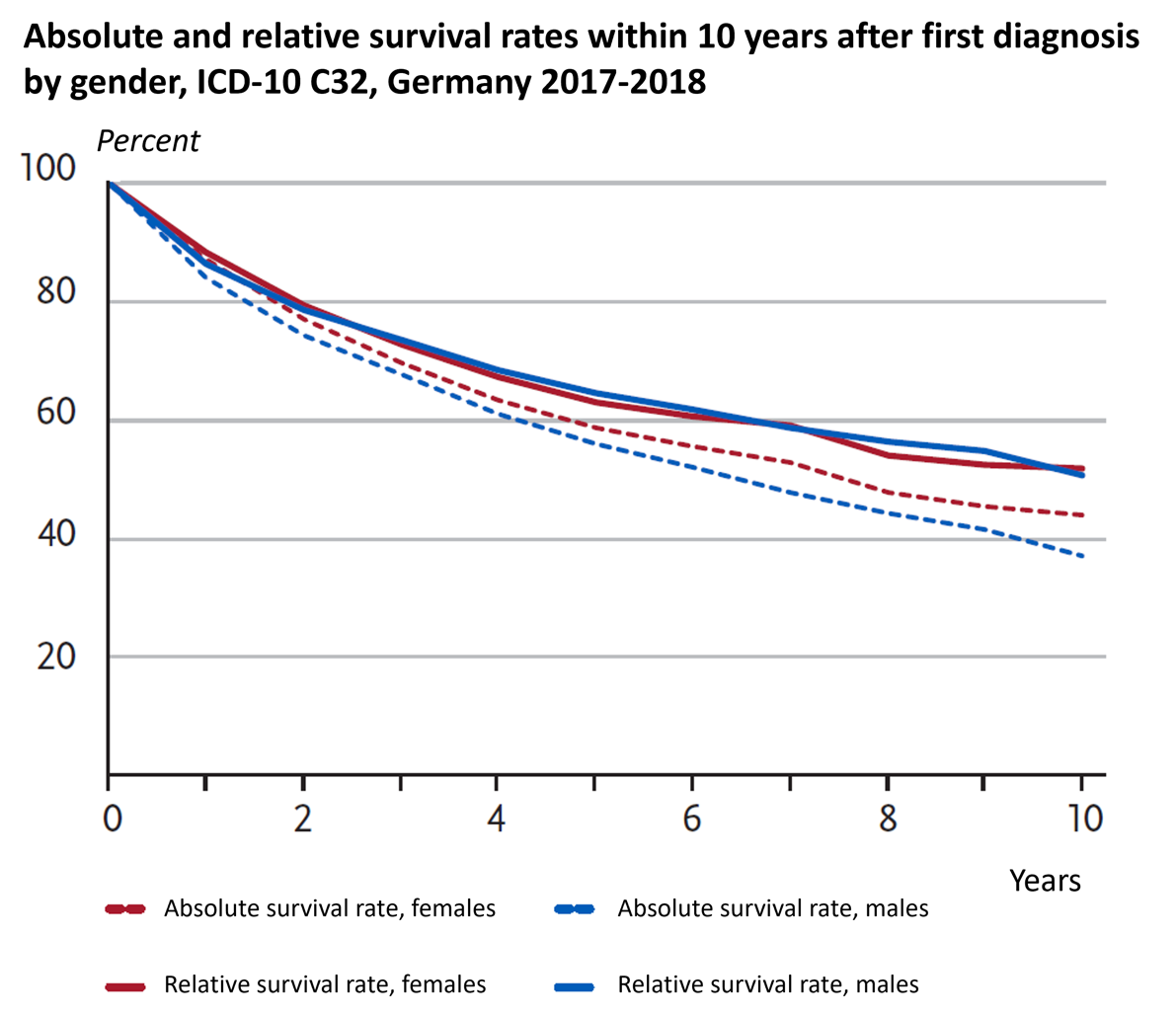
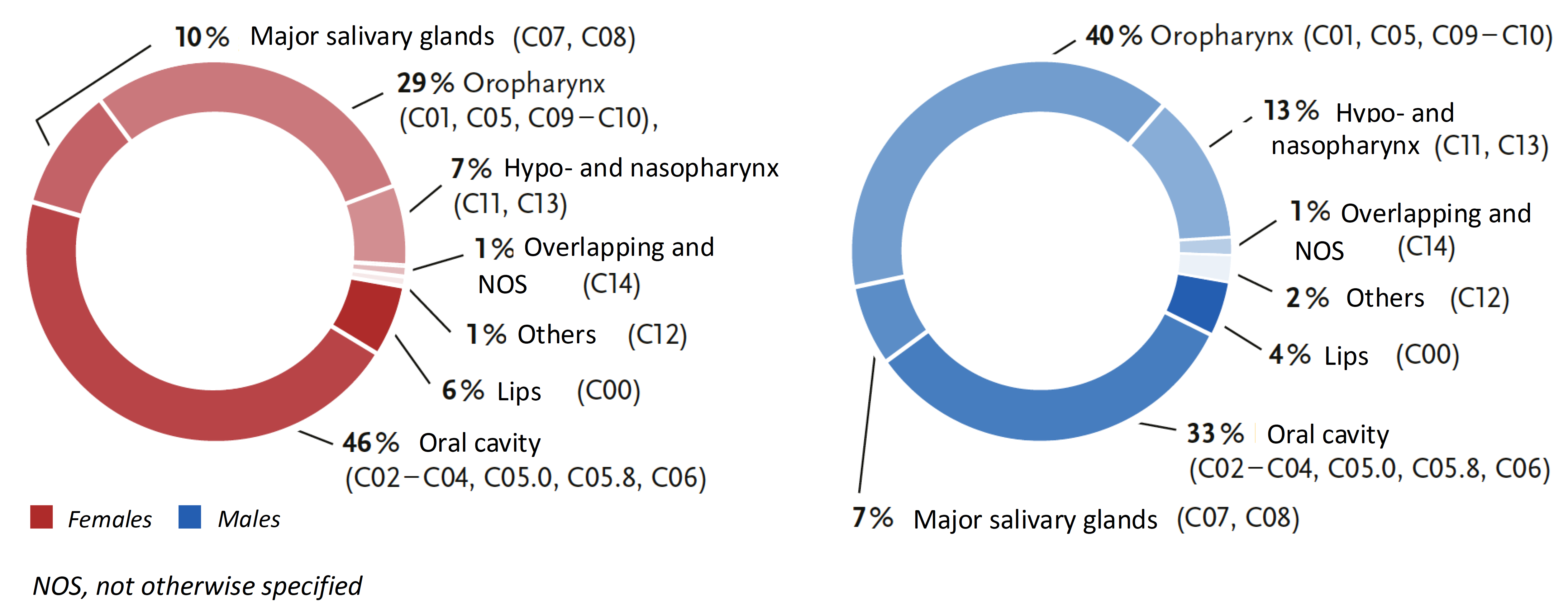
Cancers of the oral cavity and pharynx constitute a heterogeneous group of malignant neoplasms. Histologically, 85% are squamous cell carcinomas, which primarily originate in the mucous membranes of the oral cavity, nasopharynx, oropharynx, and hypopharynx. Approximately 5% of neoplasms in the oral cavity and pharynx are adenocarcinomas, which occur primarily in the salivary glands. Men are diagnosed more frequently and 2-3 years earlier than women. Age-standardized incidence rates increased for both sexes between 1999 and 2011. Since 2011, they have remained roughly constant among women, while a slight decline has been observed among men. The corresponding mortality rates have declined slightly among men over the entire period, while they have remained virtually unchanged among women. Overall, women have higher relative 5-year survival rates (63%) compared to men (54%) [1]. This is partly due to the fact that women have a lower proportion of cancers of the floor of the mouth, tongue, and pharynx caused by tobacco and alcohol use, which are associated with a lower probability of survival. According to the UICC tumor stage data currently available only for oral cavity cancers (C02-C06), just under one in three oral cavity tumors in women is diagnosed at early stage I, but only one in four cases in men.
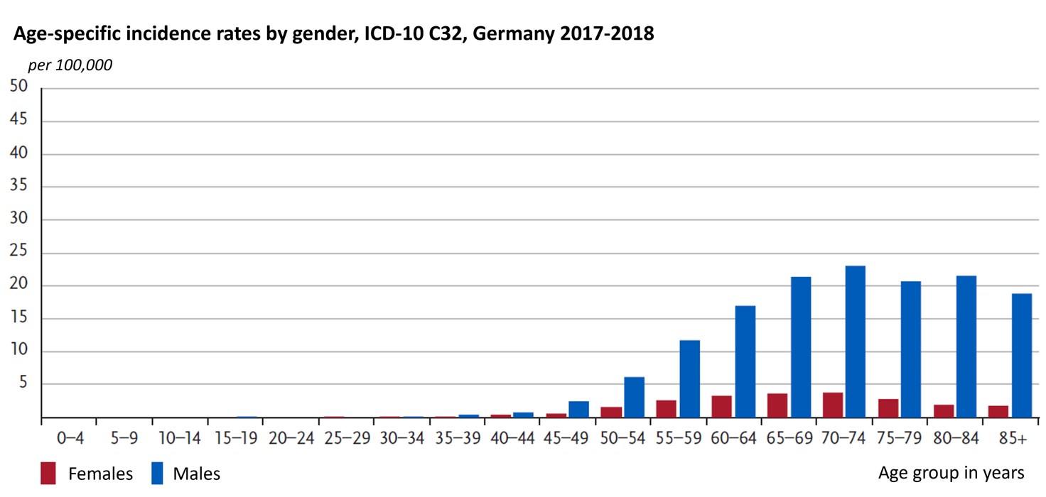
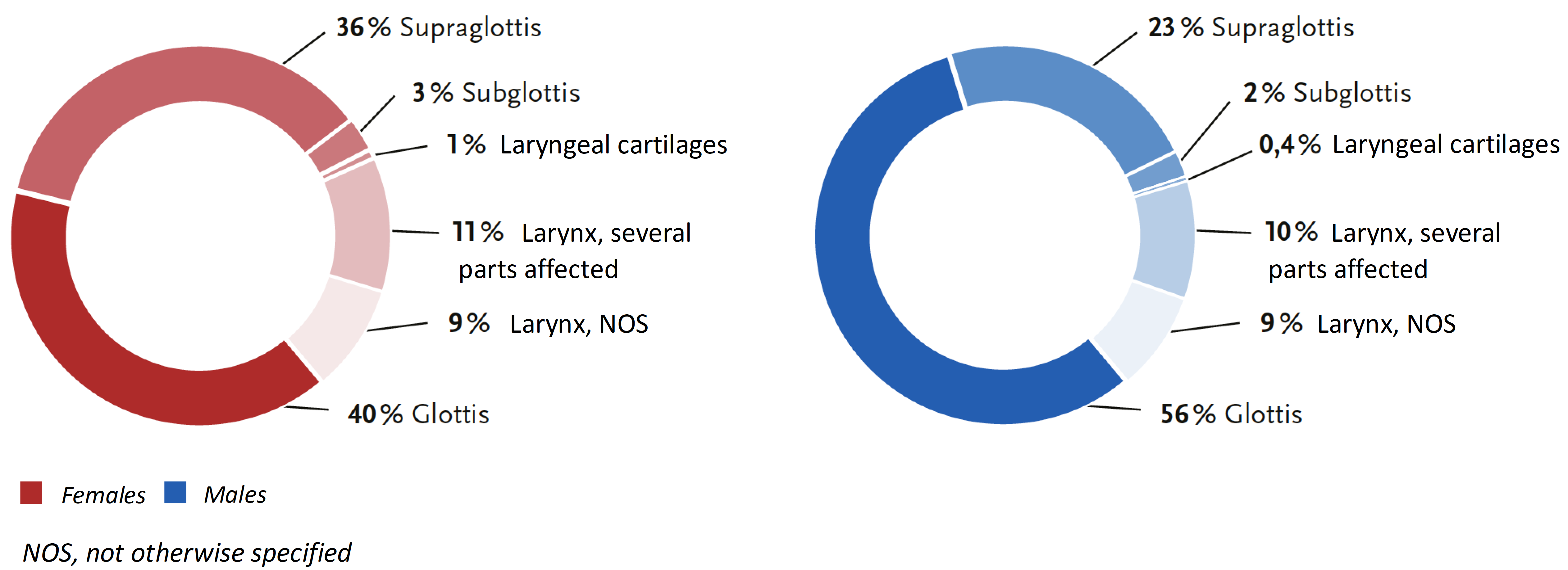
Laryngeal cancers are almost exclusively squamous cell carcinomas. Men are diagnosed with this cancer significantly more often than women: of the approximately 3,200 new cases in 2023, only about one in six affected a woman. The median age at diagnosis in 2023 was 68 years for both women and men, which is earlier than for cancer overall. Age-specific incidence rates for both women and men peak between the ages of 65 and 79. Incidence and mortality rates among men have been declining since the late 1990s. Rates among women, however, remain nearly constant. A regional comparison reveals slightly higher rates in the eastern and northern regions of Germany. The relative 5-year survival rates for women (62%) and men (66%) do not differ significantly. At 51%, a higher proportion of men are diagnosed with early tumor stages (stages I/II) than women, at 44% (according to the 8th edition of the TNM classification). Due to anti-smoking campaigns, a decline in the incidence of the disease is evident, particularly in the United States, whereas this trend is not observed in Germany due to the lack of effective programs. Smoking is associated with a poorer prognosis in patients with HPV-associated oropharyngeal carcinoma, comparable to that seen in HPV-negative carcinomas [1, 2, 3].
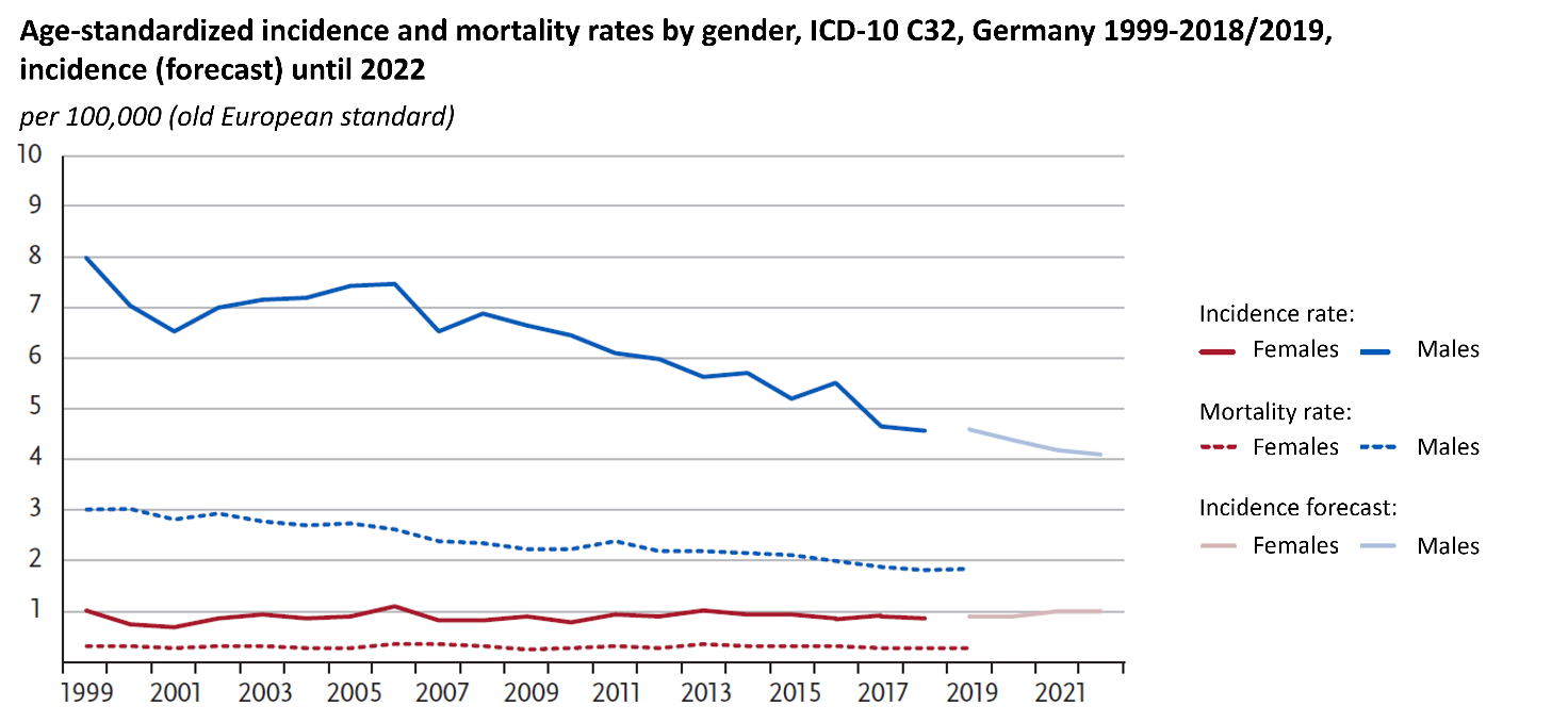
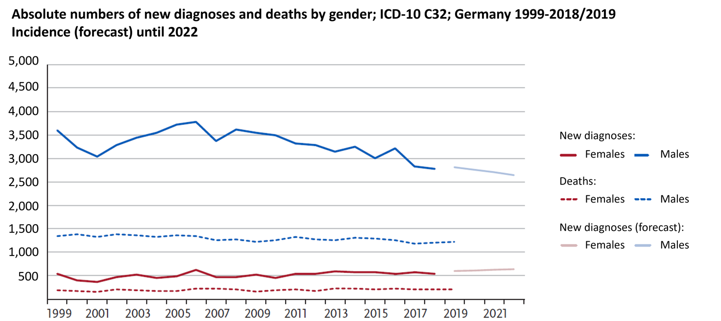
The median age at diagnosis is 63 years for men and 65 years for women, which is 7 years (men) and 4 years (women), below the median age at diagnosis for cancer overall. The median age at death for the most common sites - the oral cavity and pharynx - is 66 years (men) and 72 years (women). Most cases of oral cavity and pharynx cancer occur in men aged 60 to 70, while in women, incidence remains constant from age 55 onward, with a slight peak among those over 85 years of age.
2.3Pathogenesis
Invasive squamous cell carcinomas in the head and neck region develop primarily through two main pathways: on the one hand, these tumors arise in association with tobacco and alcohol use, and on the other hand, through infection with human papillomaviruses, particularly HPV-16. Tumors of the head and neck region develop through a complex, stepwise process involving the accumulation of genetic alterations. In particular, the inactivation of tumor suppressor genes and the activation of proto-oncogenes play a role, ultimately leading to genetic instability. While carcinogenesis for substance-associated tumors is a multifaceted and, in individual cases, complex process, the development of HPV-associated carcinomas follows a specific pattern: the expression of the viral proteins E6 and E7 leads to the inactivation of p53 and Rb [4].
2.4Risk Factors
The risk of developing head and neck squamous cell carcinoma (HNSCC) is increased by the following factors:
Alcohol consumption [5]
HPV (especially in the oropharynx) [9]
Poor oral hygiene [10]
Chronic mechanical irritation [12]
Betel nut (areca nut) consumption (oral cancer) [13]
Positive family history of head and neck tumors [14]
Long-term immunosuppression [15]
Rare: Fanconi anemia, Li-Fraumeni syndrome, Bloom syndrome, ataxia telangiectatica, congenital dyskeratosis, lichen ruber planus.
Smoking and regular alcohol consumption, in particular, are by far the most important risk factors, as they potentiate each other’s effects [16].
3[Chapter not relevant]
4Clinical Characteristics
Symptoms of HNSCC depend on the location of the tumor. Clinical signs such as weight loss, pain, dysphagia, odynophagia, and hemoptysis may occur across all locations.
Clinical symptoms by tumor location:
Oral cavity: non-healing ulcers, loose teeth, dysarthria as a sign of invasion of deep muscle layers.
Oropharynx: sleep apnea syndrome, neck swelling, globus sensation, dysphagia, odynophagia, aspiration.
Hypopharynx: dysphagia, odynophagia, aspiration, otalgia due to involvement of cranial nerves V, VII, IX, and X.
Larynx: Hoarseness, stridor, dysphagia, odynophagia, aspiration.
5Diagnosis
5.1Diagnostic Procedures
In addition to a detailed medical history, the standard diagnostic approach includes a combination of inspection, palpation, and endoscopic examination or flexible laryngoscopy. Furthermore, performance status (ECOG, Karnofsky), nutritional status, psychosocial history, dental status, and speech and swallowing function should be evaluated. For patients over 70 years of age, a geriatric assessment is also recommended. A panendoscopy (endoscopy of the pharynx and larynx, the trachea and upper bronchi, as well as the esophagus) under anesthesia is an important component of staging examinations for tumors of the larynx and pharynx and serves to confirm the histological diagnosis, accurately assess tumor size prior to definitive therapy, and rule out synchronous secondary malignancies.
A CT or MRI of the neck should be performed to rule out metastases and assess resectability. Particularly in cases of advanced tumors, expanded diagnostic imaging via CT of the thorax, and abdomen is recommended to detect any distant metastases (or secondary malignancies). CT is superior to MRI in detecting lymph node metastases, while MRI is better at visualizing perineural tumor infiltration, cartilage infiltration, and intracranial infiltrations. The two modalities should therefore be considered complementary.
In cases of suspected cervical lymph node metastasis and an undetectable primary tumor (cervical CUP syndrome), a fine-needle biopsy of the affected lymph nodes (LNs) should be performed. Ultrasound, as an examiner-dependent method, is equivalent to CT or MRI for evaluating the soft tissues of the neck. Positron emission tomography with CT (PET-CT) is superior to CT in detecting occult lymph node metastases. In general, for cross-sectional imaging - and particularly for PET-CT - due to false-negative findings in metastases <5 mm, the procedure cannot be used to stratify patients for or against definitive therapy of the lymphatic drainage pathways [17].
PET-CT can detect distant metastases that would otherwise go undetected, thereby modifying the therapeutic approach for a primary tumor that otherwise appears resectable and, in particular, determining the approach for surgical removal of cervical lymph nodes. In Germany, PET-CT is reimbursed in situations where, following completion of primary chemoradiotherapy, a decision must be made as to whether cervical lymph nodes need to be removed, based on a resolution by the Joint Federal Committee [18]. Furthermore, the added value of functional imaging lies in the search for occult primary tumors in cases of cervical lymph node metastasis and in follow-up care, when it is necessary to differentiate between therapy-induced tissue changes and tumor recurrence.
5.2Histopathological Assessment
The diagnosis is usually made by biopsy of the primary tumor or, if accessible, a lymph node metastasis.
HNSCC are classified according to the current 5th edition of the WHO classification. They are divided into subtypes with prognostic significance (verrucous/basaloid/sarcomatoid/“conventional”). “Conventional” carcinomas (keratinizing/non-keratinizing) are graded according to their similarity to normal squamous epithelium (G1/G2/G3); however, it should be noted that, according to the WHO classification, the prognostic significance of this grading is very limited. Tumors of the oropharynx have a special role, as HPV testing should be performed for all of them. For this purpose, the WHO classification recommends immunohistochemical staining for p16. p16 is established as a surrogate marker for HPV positivity in the oropharynx (which includes the tonsils and the base of the tongue). A tumor from this site is considered HPV positive if more than 70% of the tumor cells show nuclear and/or cytoplasmic staining. Other testing methods for HPV typing include, for example, PCR testing or RNA detection. It should be noted that HPV-associated HNSCCs are not graded. Due to the inherently fragmented basement membrane in lymphoepithelial tissue, there are no precursor lesions (carcinoma in situ) in these carcinomas - they are always classified as invasive.
The pathological evaluation of resected tissue specimens should include pathological staging according to the current TNM/UICC classification. This includes the following parameters:
1. Tumor size (pT1-4); for oral HNSCC, the depth of invasion should also be specified.
2. Lymph node status: for oropharyngeal carcinomas, a distinction is made between p16-negative and p16-positive oropharyngeal carcinomas.
2a. Lymph node status for all head and neck carcinomas as well as for p16-negative oropharyngeal carcinomas (pN1-3), which includes the number of resected lymph nodes, the number of those affected, and their location. With regard to the lymph nodes, the size of the largest metastasis and any evidence of extracapsular extension (ECE) should be reported. A relevant change in the 9th edition of the TNM classification is the inclusion of clinically or pathologically detected extracapsular tumor growth in lymph nodes for p16-positive oropharyngeal carcinomas.
3. Grade: G1/G2/G3
4. Perineural sheath infiltration (Pn), lymphangiosis or hemangiosis carcinomatosa (L or V). These parameters are classified as present or absent (0/1).
5. Resection status (R0/R1/R2/RX): This indicates whether carcinoma cells are detectable macroscopically or microscopically at the oriented resection margins; furthermore, the width of the tumor-free resection margins must be specified.
These parameters are considered prognostically significant and are decisive for adjuvant therapy stratification. As previously mentioned, the predictive value of the grading is limited here. It should be noted that the current TNM classification classifies p16-negative and p16-positive HNSCC as separate entities within distinct systems.
p16-positive | |||
|---|---|---|---|
Stage 0 | Tis | N0 | M0 |
Stage I | T0–T2 | N0–N1 | M0 |
Stage II | T0–T2 | N2 | M0 |
T3 | N0–N2 | M0 | |
Stage III | T0–T3 | N3 | M0 |
T4 | Any N | M0 | |
Stage IV | Any T | Any N | M1 |
In addition to these mandatory parameters, it is recommended to provide information regarding the growth pattern of the carcinoma: numerous studies have now demonstrated the prognostic significance of tumor budding, so it can be assumed that this will be included in future guidelines. Tumor budding can be classified as absent, weak, or strong.
So-called “cancer of unknown primary (CUP)” cases represent a special category. These are squamous cell-differentiated lymph node metastases for which no primary tumor can be detected. In such cases, p16 immunohistochemistry and EBV in situ hybridization of the lymph node metastasis are recommended as standard. A positive result may indicate a small HPV-associated HNSCC or an EBV-associated nasopharyngeal carcinoma as the primary tumor. If no primary tumor can be identified even after these and additional tests, CUP is assigned its own classification scheme in the current UICC/TNM classification. See Onkopedia LL CUP Syndrome for more information.
PD-L1 expression status should be determined by immunohistochemistry at the time of initial diagnosis. Under the current approval status, the Combined Positive Score (CPS) is used as a predictive biomarker for the use of pembrolizumab; a score of ≥1 is classified as positive. The cutoff value applies to both the metastatic/recurrent setting and the locally advanced neoadjuvant/adjuvant setting.
6Treatment
In the treatment of locoregionally confined HNSCC, in addition to surgery, radiotherapy - alone or in combination with systemic therapy - is available as a curative treatment option. Due to the complexity of treatment options, recommendations should always be discussed and decided in a multidisciplinary setting (multidisciplinary tumor board). In very early tumor stages (T1-2 N0 M0), surgery alone or radiotherapy alone are available. The choice of treatment path depends largely on functional aspects and the patient’s ability to tolerate treatment. In addition to tumor-specific factors, patient-specific aspects play a particularly important role, as comorbidities typical of this entity - often involving potential cardiovascular, pulmonary, or hepatic impairments - significantly complicate treatment and can lead to de facto inoperability even in patients with tumors that are surgically resectable [19].
A primarily surgical approach combined with perioperative immunotherapy is recommended for T3/T4 oral cavity carcinomas. Advanced hypopharyngeal carcinomas should be treated primarily with surgery, particularly in cases of cartilage invasion. For T3/T4a tumors of the larynx and hypopharynx, the possibility of larynx-preserving surgery, with induction therapy if indicated, should be discussed within the framework of a multidisciplinary tumor board [20]. In early T stages and selected patients with advanced T stages, a transoral approach is preferred, if technically feasible, due to its lower morbidity. Techniques using classic cold instruments, laser surgery, and transoral robotic surgery are employed here. Depending on the expected tissue defect and functional impairments, reconstructive surgery is performed using regional flaps or free flaps. The standard surgical treatment for head-and-neck soft tissue is neck dissection. The extent and radicality of the procedure depend on the tumor type and the T and N stages. Salvage surgery may be indicated in cases of tumor progression during or after primary radio(chemo)therapy or in resectable recurrent tumors.
6.1Radiotherapy as Part of a Curative Treatment Approach
Techniques such as intensity-modulated radiotherapy (IMRT), volume-modulated arc therapy (VMAT), and image-guided radiation therapy (IGRT) reduce the morbidity associated with radiotherapy [69], making these techniques the standard of care. Adaptive radiotherapy (ART) is also being used increasingly. With this approach, the radiation plan can be adjusted on a daily basis as needed. Relevant at-risk organs such as the salivary glands, oral cavity, and pharynx should be spared in accordance with international standards, without falling below the recommended dose in the respective target volume.
6.1.1Definitive Radiotherapy
In the advanced stages III, IVa, and IVb, concurrent chemoradiotherapy (RCT) is the standard of care rather than radiotherapy alone. Radiotherapy should be administered using conventional fractionation at 5 × 2 Gy per week up to a target volume dose of 70 Gy in the area of the involved lymph nodes and the primary tumor, or another established regimen with a biologically equivalent total dose should be used. Irradiation of unaffected (elective) lymph node levels should be performed at 45-54 Gy with single doses of 1.5-1.8 Gy. Radiotherapy can be administered using a simultaneously integrated boost or a sequential boost.
The treatment approach of definitive radio(chemo)therapy has been studied in particular for stage III and IV tumors of the oropharynx and larynx in non-metastatic disease. In general, a reduction in the radiation dose and treatment volume should not be performed outside of clinical trials for either HPV-positive or HPV-negative tumors.
For radiation planning, in addition to contrast-enhanced CT for radiation planning, all available imaging studies (e.g., FDG-PET-CT, MRI), panendoscopy findings, and the patient’s clinical examination findings should be taken into account. The target volume is defined according to current recommendations [70, 71, 72, 73].
Numerous studies have evaluated the effectiveness of cisplatin in combination with radiotherapy. The MACH-NC meta-analysis included individual patient data from 107 randomized trials involving a total of 19,805 patients [21, 38]. Chemotherapy was administered either as induction therapy, concurrently, or as adjuvant therapy following locoregional therapy. Simultaneous RCT proved to be the most favorable approach. In the 71 randomized trials involving 10,680 patients who received simultaneous RCT, the hazard ratio (HR) for mortality decreased to 0.83 (p<0.0001) compared with radiotherapy alone, and the absolute survival benefit at 5 years was 6.5% [21]. In most studies, cisplatin was administered as monotherapy, sometimes in combination with 5-fluorouracil (5-FU), alongside radiation therapy. Cisplatin was typically administered at a dose of 100 mg/m² body surface area (BSA) three times during radiotherapy (total dose 300 mg/m2). In the only study showing negative results, cisplatin was evidently underdosed at a total of 20 mg/m² weekly × 7 [21]. In randomized trials involving patients over 70 years of age - albeit with a small patient sample size - no survival benefit from concurrent chemotherapy could be demonstrated. In contrast, large prospective databases also show a survival benefit in patients over 70 years of age if they were in good general condition (ECOG 0-1). In older patients, therefore, the indication for concurrent chemotherapy must be assessed more strictly, taking into account general condition/ECOG and comorbidities [22]. Cisplatin-based therapy, usually as monotherapy, is considered the standard of care in definitive chemoradiotherapy. For patients unsuitable for cisplatin, carboplatin/5-FU are available as alternatives. In the presence of contraindications to platinum, mitomycin C ± 5-FU or docetaxel (if HPV-negative) [74] may be considered in the definitive setting [23, 24]. In contrast to the favorable data from the Bonner Study for oropharyngeal carcinomas [24], current evidence indicates that cetuximab is inferior to cisplatin-based therapy [25, 26, 27]. Even in HPV-negative tumors, prospective databases and a meta-analysis show that cetuximab is inferior to cisplatin [28]. Induction chemotherapy may be considered as part of larynx-preserving protocols.
An analysis by the MACH-NC group once again confirmed the benefits of RCT. Given the large number of patients, the absolute survival benefit of RCT compared to radiotherapy alone was also demonstrated for the individual tumor sites. After 5 years, the absolute survival benefit following RCT was 8.9% for tumors of the oral cavity, 8.1% for the oropharynx, 5.4% for the larynx, and 4% for tumors located in the hypopharynx [29]. According to current data, weekly administration of 40 mg/m² cisplatin does not appear to be inferior to regimens with weekly administration of 100 mg/m² cisplatin, but is better tolerated; in particular, a significantly lower rate of nephrotoxicity is observed. A cumulative dose > 200 mg/m² appears to be important in this context [30, 31].
Given the favorable prognosis of HPV-positive oropharyngeal carcinomas, the question arose as to what extent de-intensification of RCT by replacing cisplatin with cetuximab is warranted. A total of three prospective randomized trials demonstrated that concurrent chemoradiotherapy with cisplatin is superior to combined therapy with cetuximab [25]. In the NRG-RTOG 1016 phase III trial, non-inferiority of cetuximab could not be demonstrated. Patients treated with radioimmunotherapy had a higher rate of locoregional relapses (17% vs. 10%) and a lower 5-year survival rate (78% vs. 85%) [32, 33]. Cetuximab should therefore be used only in patients who have a contraindication to chemotherapy. In patients who are not eligible for cisplatin, systemic therapy with carboplatin/5-FU, docetaxel, or carboplatin/mitomycin C, cetuximab may be administered [45, 77, 78, 79, 80].
The use of checkpoint inhibitors has been investigated in several phase II/III trials in combination with radiotherapy for advanced tumors. Based on these data, their use in combination with definitive radiotherapy cannot be recommended [34, 35, 36].
Preoperative radiotherapy alone, intended to improve operability and ablate microscopic tumor components outside the resection margins, has not yet gained widespread acceptance.
6.1.2Postoperative Radiotherapy
Adjuvant radiotherapy for HNSCC is indicated in the presence of the following risk factors
pT ≥ 3
An affected lymph node > 3 cm or > 1 affected lymph node
Perineural invasion (Pn1)
Lymphovascular/vascular invasion (L1/V1)
For oral cavity carcinoma, consider RT in cases of pN1 [81, 84]
Simultaneous chemoradiotherapy is indicated in the presence of the following risk factors:
Extracapsular extension (ECE+)
Tumor-free resection margin < 5 mm or R1
Adjuvant treatment should be initiated within 6 weeks after surgery, provided there are no wound healing complications.
For radiation planning, in addition to the radiation planning CT scan (if possible, with and without contrast), all available imaging studies (e.g., FDG-PET-CT, MRI, preoperative imaging), the panendoscopy findings, the surgical report (if applicable), the pathology report, and the patient’s clinical examination findings should be taken into account. The target volume is defined according to current recommendations [70, 71, 72, 73, 75, 76].
The dose volume for radiotherapy at 5 × 2 Gy per week is as follows:
Primary tumor region in cases of R1 resection/narrow resection margin and lymph node regions with ECS: 64-66 Gy
Primary tumor region and lymph node regions without ECS: 56-60 Gy
Elective lymphatic drainage areas: 50 Gy
Other established regimens with a biologically equivalent total dose may be used. A reduction in radiation dose and treatment volume should generally not be performed outside of clinical trials for either HPV-positive or HPV-negative tumors.
The extent to which the addition of cisplatin-based chemotherapy in advanced stage III or IV tumors leads to prolonged survival is controversial. Weekly administration of cisplatin at 40 mg/m² is not inferior to 3-weekly administration of 100 mg/m² [37]. Postoperative RCT resulted in improved local control compared with radiation therapy alone, as demonstrated in three large, independent studies (EORTC 22931: n = 334; RTOG 9501: n = 459; and ARO96-3, published only as an abstract: n = 440) [77]. However, after 10 years of follow-up, a pooled analysis of the EORTC and RTOG studies revealed a persistent significant difference in disease-free survival and local control only for the high-risk group of patients with extracapsular growth and a positive resection margin. For patients with an increased risk of recurrence - based on T3 and T4 tumors, perineural or vascular infiltration, and two or more involved lymph nodes - the addition of chemotherapy to radiation therapy is not supported due to the non-significant differences in disease-free survival and local control. A recent long-term analysis of the EORTC22931 and RTOG9501 studies demonstrates a survival benefit from concurrent platinum-based chemoradiotherapy and suggests that potentially more patients than those with a positive resection margin and ECE may benefit from concurrent chemoradiotherapy. Due to the increased non-tumor-specific mortality in the arm of patients receiving chemoradiotherapy, the use of combined chemoradiotherapy in the postoperative setting must be strictly evaluated while taking comorbidities and ECOG status into account [82].
6.2Adjuvant Systemic Therapy
According to a meta-analysis including a total of approximately 2,500 patients, adjuvant chemotherapy alone following successful primary therapy (R0 resection) is not indicated, as the 5-year survival rate of 48.4% was no better than that in the control arm (49.4%) [38, 39].
Data from phase III trials are available regarding the adjuvant use of checkpoint inhibitors. The NIVOPOSTOP study (GORTEC 2018-01) was a randomized, open-label phase III study of postoperative treatment in patients with resected, locally advanced HNSCC and a high risk of recurrence, defined by R1 or ECE-positive lymph nodes. The study investigated whether adding nivolumab to standard adjuvant therapy consisting of cisplatin and radiation therapy could improve disease-free survival (DFS). One dose of nivolumab was administered before chemoradiotherapy, three doses concurrently with RT, and six doses after RCT. The study primarily included patients with oral cavity carcinomas (58%). The results demonstrated a significant benefit for the experimental arm: the additional administration of nivolumab reduced the risk of relapse or death (HR 0.76; 95% confidence interval [CI] 0.60-0.98). At a median follow-up of 30.3 months, the 3-year DFS rate was 63.1% in the nivolumab arm compared with 52.5% in the control arm. Unlike other adjuvant therapies with checkpoint inhibitors, nivolumab was initiated before the start of radiation therapy. The benefit was observed regardless of PD-L1 expression and was primarily due to a reduced rate of local recurrence [64]. As of the publication date of this guideline (June 2026), nivolumab is not EMA-approved for this indication.
The Imvoke010 study showed conflicting results. In this global, randomized, double-blind, placebo-controlled study, patients with high-risk HNSCC were randomized after completing definitive multimodal therapy to receive either atezolizumab 1200 mg every 3 weeks for 1 year or placebo. The study included, among others, tumors of the oral cavity, larynx, and hypopharynx, as well as HPV-negative oropharyngeal carcinomas in stages IVa/IVb and HPV-positive oropharyngeal carcinomas in stage III. A total of 406 patients were randomized, with 203 in each arm. The primary endpoint was investigator-assessed event-free survival (EFS). After a median follow-up of 46.5 months, the median EFS was 59.5 months with atezolizumab versus 52.7 months with placebo; the difference was not statistically significant, with an HR of 0.94 (95% CI 0.70-1.26; p = 0.68). No difference was observed in overall survival either; the 24-month OS was 82.0% with atezolizumab and 79.2% with placebo [65].
6.3Systemic Therapy
6.3.1Induction Chemotherapy
In older studies, primary chemotherapy for locally advanced but non-metastatic HNSCC proved highly effective, with remission rates of ≥ 80%, although it remained unclear whether this approach leads to better long-term outcomes than simultaneous RCT. A meta-analysis of 31 studies involving 5,311 patients who received locoregional treatment either immediately or only after induction chemotherapy (ICT) demonstrated a clinically insignificant increase in the 5-year survival rate from 30% to 32.4% for induction therapy [21]. The comparison between induction and concurrent chemoradiotherapy also points to the greater effectiveness of concurrent chemoradiotherapy.
Two meta-analyses evaluating individual data from 33 randomized trials involving a total of 5,211 patients demonstrated that induction therapy in patients with HNSCC reduces the rate of distant metastasis by 8%, but had no effect on either local control or overall survival [40, 41]. To preserve the larynx in cases of very advanced tumors that would otherwise require laryngectomy or pharyngectomy, induction chemotherapy followed by radiotherapy alone can be used. Larynx preservation rates are higher when induction therapy is followed by chemoradiotherapy, although this is associated with increased toxicity and comparable survival rates [42]. The randomized TAX323 trial investigated the superiority of a combination of docetaxel, cisplatin, and 5-FU (TPF) in induction therapy compared to the two-drug combination of cisplatin and 5-FU (PF). The trial demonstrated a survival benefit in favor of TPF, albeit with high toxicity [43]. A follow-up examination after the first cycle using imaging and panendoscopy allows for an assessment of the likelihood of larynx preservation, whereby a tumor size reduction of at least 30% should be achieved to justify continuing induction chemotherapy for a total of 3 cycles [44]. Patients with cartilage infiltration of the larynx should be offered a primarily surgical approach. In other locations outside the larynx, ICT currently has no role [45].
6.3.2Perioperative Immunotherapy
The KEYNOTE-689 study was an international, randomized, open-label phase III trial investigating a perioperative immunotherapy regimen in patients with newly diagnosed, resectable, locally advanced HNSCC at stages III and IVa. The study compared standard therapy - consisting of surgery and postoperative radiotherapy with or without cisplatin - with an experimental approach in which pembrolizumab was added to standard treatment both neoadjuvantly and adjuvantly. In the experimental arm, patients received two cycles of pembrolizumab prior to surgery, followed by 15 doses of pembrolizumab after resection, in addition to standard adjuvant therapy. The primary endpoint was event-free survival (EFS). At a median follow-up of 38.3 months, a significant benefit was observed in favor of the pembrolizumab arm. In the overall population, the 36-month EFS rate was 57.6% in the pembrolizumab group compared with 46.4% in the control arm; corresponding to a hazard ratio (HR) of 0.73 (95% CI 0.58-0.92; p = 0.008). The median EFS in the overall population was 51.8 months in the pembrolizumab arm compared with 30.5 months in the standard-of-care arm. There was no difference in the rate of local recurrence between the two treatment arms. However, in the pembrolizumab group, the rate of distant metastasis was significantly reduced. Of clinical relevance was the fact that the feasibility of surgery was not significantly impaired by neoadjuvant pembrolizumab administration [66]. The proportion of patients with a major pathological response (mPR) and complete pathological response (pCR) in the CPS ≥1 population was 9.8% and 3.2%, respectively. Although recent analyses of the Keynote-689 study suggest an impressive EFS in this small population, a de-escalation of postoperative therapy is not currently recommended outside of clinical trials.
6.3.3First-line Palliative Therapy
In the presence of distant metastases or locoregionally advanced disease that can no longer be controlled by surgery or radiotherapy, palliative systemic therapy should be offered if the patient’s general condition is good (ECOG 0-2). Median overall survival with palliative systemic therapy ranges between 12 and 15 months [46, 47, 48]. In cases of oligometastatic disease, the option of surgery or radiotherapy - either in addition to or instead of systemic therapy - should be discussed in a multidisciplinary tumor board.
In most cases, chemotherapy results in partial remission of the tumor. Complete remissions are rare. In addition to prolonging survival, the goal of therapy is to maintain or improve quality of life.
The standard of care for first-line palliative therapy has been established by the Keynote-048 study and the TPExtreme study. In the Keynote-048 study, the long-standing standard of care - the EXTREME protocol with cisplatin, 5-FU, and cetuximab (PF-C) followed by cetuximab maintenance [43] - was randomly compared against the checkpoint inhibitor pembrolizumab alone or against the combination of pembrolizumab, cisplatin, and 5-FU (PF-PEM) followed by pembrolizumab maintenance [46]. As a monotherapy, pembrolizumab was able to prolong overall survival to 14.9 versus 10.7 months, particularly in tumors with high PD-L1 expression (CPS ≥ 20), with a significantly better side effect profile. However, the response rate (23.3% vs. 36.1%) is lower than with PF-C, and there are also more patients with pembrolizumab monotherapy who experience primary progression than with PF-C. For patients with a CPS ≥ 1, there is also a benefit in overall survival at 12.3 months versus 10.3 months, although 38.9% of patients in this group experience primary progression. With PF-PEM, overall survival is also significantly prolonged compared to PF-C in patients with CPS ≥ 1, with a comparable remission rate. However, the rate of adverse effects is comparable to that observed in the EXTREME trial and significantly higher than with pembrolizumab alone. Currently, there are no data available on the use of immunotherapy in a palliative setting following prior neoadjuvant/adjuvant therapy from which a treatment recommendation can be derived.
Regarding chemotherapy in combination with the EGFR antibody cetuximab, a randomized phase II study compared PF-C with the TPEx protocol (cisplatin, docetaxel, cetuximab), each followed by cetuximab maintenance [47]. In the TPEx arm, chemotherapy was shortened from 6 cycles to 4 cycles, and the total cisplatin dose was reduced by 50% compared to PF-C. Overall survival was comparable, with lower overall toxicity and a shorter duration of chemotherapy in the TPEx arm. The response rate with the docetaxel-containing regimen was also comparable to that of the control arm at 57%, demonstrating the high efficacy of the regimen. Thus, TPEx can be used as an alternative to PF-C as a first-line therapy in patients in good general condition, under remission pressure, and with a high tumor burden, regardless of PD-L1 status. G-CSF administration was mandatory as part of the study treatment and is recommended for routine clinical practice.
For patients under high remission pressure or with CPS 0 who are not suitable for intensive therapy as per the TPEx protocol, retrospective data are available on the weekly administration of paclitaxel, carboplatin, and cetuximab. 52% of patients had a reduced ECOG performance status of 2, and a response rate of 43.3% with disease control of 65% was achieved. The median progression-free survival was 5.8 months, and the median overall survival was 11.7 months. Given that these are retrospective data, this combination is an option for patients with contraindications to TPEx [83].
1 CPS = Combined Positive Score;
2 Performance status according to ECOG (Eastern Cooperative Oncology Group)
With regard to the situation of a relapse while undergoing adjuvant immunotherapy, there are currently no reliable data available on the therapeutic approach. The authors of the guideline recommend an approach analogous to second-line palliative therapy following immunotherapy failure.
6.3.4Second-line Palliative Therapy
For patients with disease progression following platinum-based chemotherapy, the checkpoint inhibitor nivolumab significantly prolonged survival to 7.5 vs. 5.1 months compared to monotherapy with a taxane, MTX, or cetuximab [49]. Nivolumab is approved for use in patients with disease progression following platinum-based therapy, regardless of PD-L1 status. In a comparable study design, similar results were achieved for the PD-1 inhibitor pembrolizumab, although statistical significance was reached only in the patient group with a PD-L1 TPS ≥ 50% [50]. For patients with a PD-L1 TPS ≥ 50%, median overall survival was significantly better with pembrolizumab (11.6 months) compared to taxane, MTX, or cetuximab (6.6 months), leading to approval for patients with a TPS ≥ 50% who have progressed after prior platinum-based therapy.
Accordingly, in the absence of contraindications to a checkpoint inhibitor, second-line therapy following P-FC or TPEx should consist of treatment with nivolumab (regardless of PD-L1 expression) or pembrolizumab (TPS ≥ 50%).
Following first-line therapy with pembrolizumab monotherapy or PF-PEM, there is no established, internationally recognized standard of care. Various retrospective analyses have demonstrated high remission rates of up to 70% with salvage chemotherapy following checkpoint inhibition [51, 67]. Prospective data on the use of paclitaxel in combination with cetuximab are now available. The study included patients who had progressed following pembrolizumab-based first-line therapy. Treatment consisted of paclitaxel 175 mg/m² every 21 days in combination with weekly cetuximab 250 mg/m² for up to six cycles, followed by cetuximab maintenance therapy. The primary endpoint was the objective response rate (ORR) at 12 weeks. The study demonstrated clinically relevant activity of the combination therapy: the ORR at 12 weeks was 43.9%, and the best objective response rate was 47.4%. The disease control rate (DCR) was 71.9%. The median progression-free survival (PFS) was 5.9 months, and the median overall survival (OS) was 12.2 months. The 6-month rates for PFS and OS were 49.0% and 73.0%, respectively. Thus, the study provided prospective evidence that the combination of paclitaxel and cetuximab exhibits significant antitumor activity following failure of pembrolizumab-based first-line therapy [68].
1 for PD-L1 TPS ≥50%;
2 off-label when cetuximab is administered without platinum
6.3.5Agents for Systemic Cancer Therapy (in alphabetical order)
6.3.5.1-Fluorouracil
5-Fluorouracil is used in systemic tumor therapy for patients with head and neck tumors in both neoadjuvant and metastatic therapy. In contrast to the therapeutic indications for 5-FU in other tumor types, it is not combined with folinic acid to enhance efficacy in HNSCC. Oral alternatives, such as capecitabine, can be used as monotherapy in advanced stages of treatment for head and neck cancers. Severe side effects include diarrhea and stomatitis. Patients with functionally relevant polymorphisms in the genes involved in 5-FU metabolism have an increased risk of severe side effects, including neutropenia and neutropenic fever. Since April 2020, it has been mandatory, in accordance with the recommendations of the European Medicines Agency (EMA), to test all patients for dihydropyrimidine dehydrogenase (DPD) deficiency (either by measuring uracil levels or by testing for the presence of specific polymorphisms) prior to starting treatment with 5-FU, and appropriate therapeutic measures must be implemented based on the results.
6.3.5.2Carboplatin
Carboplatin can be used as an alternative in cases where cisplatin is contraindicated, particularly when nephrotoxicity is a problem. It has not yet been compared in a randomized trial against cisplatin, and cisplatin remains the standard of care in combination with radiotherapy and in palliative systemic therapy. For fit patients aged ≥ 70 years (geriatric assessment recommended), the combination of carboplatin, 5-FU, and cetuximab demonstrated a PFS of 7.2 months and an OS of 14.7 months; thus, for fit older patients receiving platinum, 5-FU, and cetuximab, the use of carboplatin may be considered [52].
6.3.5.3Cetuximab
Cetuximab competes with ligands for the binding site on the EGF receptor. If cetuximab binds, phosphorylation of the tyrosine kinase and activation of the signaling cascade are prevented. Furthermore, the receptor is internalized and subsequently degraded, resulting in a decrease in EGFR expression. A third mechanism, antibody-dependent cellular cytotoxicity (ADCC), leads to the migration of NK and cytotoxic T cells, resulting in the lysis of antibody-bearing cells. An additional antiproliferative effect is achieved through the reduced release of angiogenic growth factors. The antibody was initially investigated in the “Bonner” study in combination with radiotherapy in a curative setting. The study demonstrated significantly longer progression-free survival (24.4 vs. 14.9 months) and overall survival (49 vs. 29.3 months) compared to radiotherapy alone [53]. In the subsequent RTOG 0522 study, cetuximab in combination with cisplatin and radiotherapy was compared with cisplatin and radiation therapy alone. No benefit was observed for the addition of cetuximab in any of the relevant endpoints [54]. The EXTREME study evaluated cetuximab in combination with chemotherapy in the palliative setting. The combination of platinum, 5-FU, and cetuximab resulted in significantly longer survival and thus became the new standard of care in the palliative setting [48]. Cetuximab was thus the first approved targeted therapy for the treatment of HNSCC.
6.3.5.4Cisplatin
Cisplatin is the standard of care in combination with radiotherapy for locally advanced disease and is used in combination therapy in the palliative setting. As part of definitive chemoradiotherapy, cisplatin is administered as monotherapy and should reach a total dose of ≥ 200 mg/m². In a phase II/III study, weekly administration of 40 mg/m² demonstrated non-inferiority in overall survival and a better side-effect profile compared to administration of 100 mg/m² every 3 weeks [55]. The fractionated administration of weekly cisplatin also demonstrated non-inferiority in the adjuvant setting [37]. The extent to which these data will replace the current standard of 100 mg/m² in both the curative and adjuvant settings is currently the subject of controversy. In palliative therapy, cisplatin is combined with 5-FU and pembrolizumab or with cetuximab plus 5-FU or docetaxel, yielding remission rates between 36% and 59% with a median overall survival of 13-14 months. Specific severe side effects (grade 3 or 4) include nausea and vomiting, nephrotoxicity, polyneuropathy, ototoxicity, hematotoxicity, electrolyte dysbalances, cardiotoxicity, and diarrhea.
6.3.5.5Docetaxel
As a monotherapy, docetaxel is among the agents with the highest activity against HNSCC, alongside platinum derivatives. In studies, doses of 40 mg/m² weekly or 100 mg/m² every 3 weeks were investigated as monotherapy in patients with HNSCC. Objective response rates ranged from 27% to 32% in previously treated patients [56, 57, 58]. In the TPEx study, docetaxel was combined with cisplatin and cetuximab. Based on efficacy data and its favorable toxicity profile compared to cisplatin, 5-FU, and cetuximab (EXTREME protocol), the docetaxel combination is considered the standard of care in first-line palliative treatment for fit patients who do not express PD-L1 [47]. Furthermore, the drug is effective as monotherapy in second-line treatment or in combination with the EGFR antibody cetuximab. Severe side effects (grade 3 or 4) include infections, nail changes, taste disturbances, stomatitis, and diarrhea. Treatment-related side effects (grade 2) include alopecia. Particularly burdensome is polyneuropathy, which may be irreversible in some cases. Common side effects such as nausea/vomiting and allergic reactions can be prevented with adequate supportive care; see Onkopedia Antiemetics.
6.3.5.6Methotrexate
Methotrexate can be used as monotherapy when platinum-based combination chemotherapy is contraindicated; in a randomized phase III trial, weekly administration of 40 mg/m² achieved a remission rate of 3.9% compared to gefitinib, with a median overall survival of 6.7 months [59]. The most common side effects of MTX were mucositis, nausea, and constipation.
6.3.5.7Mitomycin C
Mitomycin belongs to the group of alkylating antibiotics, which exert an antiproliferative effect. In the ARO 95-06 study, mitomycin was administered in combination with 5-FU and hyperfractionated radiotherapy and compared with radiotherapy alone. After a median follow-up period of 8.7 years, local tumor control was 12% higher than the control rate for radiation therapy alone. Mitomycin C/5-FU can therefore be used in combination with radiotherapy as an alternative to cisplatin or carboplatin in platin-ineligible patients [23].
6.3.5.8Nivolumab
Nivolumab is a checkpoint inhibitor that binds to PD-1 and has been evaluated in patients with previously treated HNSCC. The CheckMate-141 phase III trial compared nivolumab with investigator’s choice (methotrexate, docetaxel, or cetuximab) in 361 platinum-refractory patients. Overall survival was 7.7 months with nivolumab compared with 5.1 months, and the 1-year survival rate was 34% versus 19.7% in the control arm. The objective response rate (ORR) was 13.3% versus 5.8%. In a preplanned exploratory analysis, overall survival was found to be dependent on PD-L1 expression ≥ 1 (8.7 versus 4.6 months). The survival benefit could not be demonstrated compared to patients who showed no PD-L1 expression [49]. Quality-of-life studies showed that no decline occurred under nivolumab compared to the control arm, in which no significant decline was recorded in 8 of 15 relevant criteria [60]. Based on the study results, the drug was approved in Europe for platinum-pretreated patients with HNSCC.
6.3.5.9Paclitaxel/nab-paclitaxel
Like docetaxel, paclitaxel is one of the active agents for HNSCC. Paclitaxel was investigated in combination with platinum and cetuximab in first-line palliative care and also in the second-line setting. The CSPOR-HN02 and CETMET studies examined the role of paclitaxel in first-line palliative therapy. Both studies included only a small number of patients. Efficacy data were comparable to those of other combinations using 5-FU or docetaxel in combination with platinum derivatives. Prospective data are now available on the use of paclitaxel in combination with cetuximab following failure of checkpoint inhibitor therapy. The study included patients whose disease had progressed following pembrolizumab-based first-line therapy. Treatment consisted of paclitaxel 175 mg/m² every 21 days in combination with weekly cetuximab 250 mg/m² for up to six cycles, followed by cetuximab maintenance therapy. The primary endpoint was the objective response rate (ORR) at 12 weeks. The study demonstrated clinically relevant activity of the combination therapy: the ORR at 12 weeks was 43.9%, and the best objective response rate was 47.4%. The disease control rate (DCR) was 71.9%. The median PFS was 5.9 months, and the median OS was 12.2 months. The 6-month rates for PFS and OS were 49.0% and 73.0%, respectively. Thus, the study provided prospective evidence that the combination of paclitaxel and cetuximab exhibits significant antitumor activity following failure of pembrolizumab-based first-line therapy [68] To date, only a small phase II study (CACTUX) has been conducted on cremophor-free albumin-bound nab-paclitaxel, which demonstrated a high ORR of 63%, a PFS of 6.8 months, and an OS of 18.8 months [61].
6.3.5.10Pembrolizumab
Pembrolizumab is a checkpoint inhibitor that binds to PD-1 and has been investigated in first-line palliative therapy as part of the Keynote-048 study, in advanced disease following platinum-based first-line palliative therapy (Keynote-040), and in the locally advanced curative setting (Keynote-689). In the Keynote-048 trial (882 patients, phase III), the combination of pembrolizumab with cisplatin and 5-FU was compared to the standard of care - cisplatin, 5-FU, and cetuximab (PFC; EXTREME regimen). Furthermore, pembrolizumab was compared as monotherapy against PFC. A comparison of the two arms with the checkpoint inhibitor was not conducted in this 3-arm study. PD-L1 expression was determined using CPS.
The study demonstrated prolonged OS compared to the control arm with chemotherapy (median 13.0 vs. 10.7 months; 2-year survival 29% vs. 19%; HR 0.77; 95% CI 0.63-0.93). The benefit of adding immunotherapy to chemotherapy was particularly evident in the PD-L1-positive population: CPS ≥20 (median OS 14.7 vs. 11.0 months, 2-year OS 35% vs. 19%, HR 0.60; 95% CI 0.45-0.82) and for patients whose tumor had a CPS ≥ 1 (median 13.6 vs. 10.4 months, 2-year OS 31% vs. 17%, HR 0.65, 95% CI 0.53-0.80).
Monotherapy compared with the PFC chemotherapy combination also showed prolonged OS, but a lower response rate (17% vs. 36%) and shorter PFS (2.3 vs. 5.2 months, HR 1.34; 95% CI 1.13-1.59). CPS ≥20 (median 14.9 vs. 10.7 months, 2-year OS 38% vs. 22%, HR 0.61; 95% CI 0.45-0.83) and for patients with CPS ≥1 (median 12.3 vs. 10.3 months, 2-year OS 30% vs. 19%, HR 0.78; 95% CI 0.64-0.96).
The results of the Keynote-048 study led to the approval of pembrolizumab, with or without chemotherapy, as first-line treatment for patients with a CPS ≥1.
Pembrolizumab had previously been evaluated in a patient population with platinum-refractory head and neck tumors against “investigator’s choice” (cetuximab, docetaxel, or methotrexate) in the KEYNOTE-040 study. Pembrolizumab prolonged survival (1-year OS 37% vs. 27%, median OS 8.4 vs. 6.9 months, HR 0.80; 95% CI 0.65-0.98). The survival benefit was particularly pronounced in tumors with high PD-L1 expression (HR 0.53; 95% CI 0.35-0.81). PD-L1 expression was determined using TPS. Based on these data, pembrolizumab was approved for pretreated patients with a PD-L1 TPS ≥50% [46, 50].
In addition, following the publication of the results of the Keynote-689 study, pembrolizumab has been approved as monotherapy for neoadjuvant treatment and subsequently for adjuvant treatment in combination with radiation therapy, with or without concomitant cisplatin therapy, and then as monotherapy for resectable locally advanced HNSCC in adults with PD-L1-expressing tumors (CPS ≥1). The KEYNOTE-689 study was an international, open-label, randomized phase III trial designed to evaluate a perioperative immunotherapy regimen in patients with newly diagnosed, resectable, locally advanced HNSCC at stages III and IVa. The study compared standard therapy - consisting of surgery and postoperative radiation therapy with or without cisplatin - with an experimental approach in which pembrolizumab was added to standard treatment both neoadjuvantly and adjuvantly. In the experimental arm, patients received two cycles of pembrolizumab prior to surgery, followed by 15 doses of pembrolizumab after resection, in addition to standard adjuvant therapy. The primary endpoint was event-free survival (EFS). At a median follow-up of 38.3 months, a significant benefit was observed in favor of the pembrolizumab arm. In the overall population, the 36-month EFS rate was 57.6% in the pembrolizumab group compared with 46.4% in the control arm; corresponding to an HR of 0.73 (95% CI 0.58-0.92; p = 0.008). The median EFS in the overall population was 51.8 months in the pembrolizumab arm compared with 30.5 months in the standard-of-care arm. There was no difference in the rate of local recurrence between the two treatment arms. However, in the pembrolizumab group, the rate of distant metastasis was significantly reduced. Of clinical relevance was the fact that the feasibility of surgery was not significantly impaired by neoadjuvant pembrolizumab administration [66].
7Bone Metastases
Bone metastases occur with a frequency of 2-22%, depending on the primary tumor site, and lead to bone fractures, spinal canal compression, or hypercalcemia in 27% of cases. Hypercalcemia, caused by the frequent secretion of parathyroid hormone-related peptide (PTHrP) in HNSCC, is the most common complication. Patients with bone metastases have a very poor median survival of only 6 months, although the use of radiotherapy and bisphosphonates has a favorable effect on survival. It is therefore recommended to initiate antiresorptive therapy upon detection of bone metastases and to combine it with radiotherapy in clinically symptomatic patients [62].
8Rehabilitation
Head and neck tumors themselves, as well as their treatments - including surgery, systemic therapy, and/or radiation therapy - often lead to significant distress, a loss of quality of life, and functional and somatic sequelae such as post-treatment chewing, speech, and swallowing disorders; radiation-induced xerostomia; weight loss ranging up to tumor cachexia; chemotherapy-induced polyneuropathy, and general weakness, up to (chronic) fatigue syndrome. As a result of these side effects, associated comorbidities (e.g., alcohol abuse), and the oncological diagnosis itself, patients experience significant psychological distress and, accordingly, require concurrent psycho-oncological treatment and psychosocial support. Therefore, targeted rehabilitation measures are necessary. These should be initiated as soon as possible after completion of primary therapy. During rehabilitation, in addition to general therapeutic services (exercise therapy, physical therapy, and occupational therapy), comprehensive nutritional counseling and nutritional support should be provided; patients should be offered a teaching kitchen; and there should be the option to administer all scientifically recognized forms of nutrition - ranging from a normal, full diet and enteral nutrition to complete parenteral nutrition. The costs of dental rehabilitation with dental implants following treatment for head and neck cancer are usually covered by statutory health insurance (Section 28 of Book V of the German SGB V). Rehabilitation facilities should be able to continue systemic tumor therapy if indicated. Patients who have not yet reached the statutory retirement age should be informed, as part of medically and vocationally oriented rehabilitation (MBOR), about services designed to facilitate their participation in working life. Other socio-medical issues, as well as any necessary patient care, should be addressed during rehabilitation. Furthermore, all patients should be offered psycho-oncological care.
9Monitoring and Follow-up
Curative treatment is followed by structured monitoring care to detect early relapses or secondary malignancies and long-term toxicities. The majority of relapses occur within the first two years after initial therapy. Clinical follow-up examinations are conducted every 3 months during years 1 and 2. Between years 3 and 5, follow-up examinations are conducted every 6 months. For locally advanced tumors, cross-sectional imaging is recommended to monitor the local findings and detect possible secondary tumors; a CT or MRI is recommended every 6 months during the first two years and subsequently every 12 months until the fifth year. PET-CT is (still) reserved for specific clinical questions and for patients who have undergone curative RCT, in order to identify positive lymph nodes for neck dissection. In addition, cross-sectional imaging should be performed in cases of clinical symptoms or abnormal clinical findings [63].
10References
From: Cancer in Germany, as of March 2, 2026. https://www.krebsdaten.de/krebs/de/content/publikationen/krebs_in_deutschland/kid_2025/kid_2025_c00_c14_mundhoehle_rachen and https://www.krebsdaten.de/krebs/de/content/publications/cancer_in_germany/kid_2025/kid_2025_c32_larynx (Accessed March 21, 2026)
Tinhofer I, Johrens K, Keilholz U et al. Contribution of human papillomavirus to the incidence of squamous cell carcinoma of the head and neck in a European population with high smoking prevalence. Eur J Cancer 2015;51:514-521. DOI:10.1016/j.ejca.2014.12.018
Gillison ML, Zhang Q, Jordan R et al. Tobacco smoking and increased risk of death and progression for patients with p16-positive and p16-negative oropharyngeal cancer. J Clin Oncol 2012;30:2102-2111. DOI:10.1200/JCO.2011.38.4099
Leemans CR, Snijders PJF, Brakenhoff RH. The molecular landscape of head and neck cancer. Nat Rev Cancer 2018;18:269-282. DOI:10.1038/nrc.2018.11
De Stefani E, Boffetta P, Oreggia F, Fierro L, Mendilaharsu M. Hard liquor consumption is associated with a higher risk of oral and pharyngeal cancer than wine consumption. A case-control study in Uruguay. Oral Oncol 1998;34:99-104. DOI:10.1016/s1368-8375(97)00062-6
Wyss A, Hashibe M, Chuang SC et al. Cigarette, cigar, and pipe smoking and the risk of head and neck cancers: a pooled analysis by the International Head and Neck Cancer Epidemiology Consortium. Am J Epidemiol 2013;178:679-690. DOI:10.1093/aje/kwt029
Blot WJ, McLaughlin JK, Winn DM et al. Smoking and drinking in relation to oral and pharyngeal cancer. Cancer Res 1988;48:3282-3287. PMID:3365707
Spitz MR. Epidemiology and risk factors for head and neck cancer. Semin Oncol 1994;21:281-288. PMID:8209260
Chaturvedi AK, Engels EA, Pfeiffer RM et al. Human papillomavirus and rising oropharyngeal cancer incidence in the United States. J Clin Oncol 2011;29:4294-4301. DOI:10.1200/JCO.2011.36.4596
Hashim D, Sartori S, Brennan P et al. The role of oral hygiene in head and neck cancer: results from the International Head and Neck Cancer Epidemiology (INHANCE) consortium. Ann Oncol 2016;27:1619-1625. DOI:10.1093/annonc/mdw224
Mahale P, Sturgis EM, Tweardy DJ, Ariza-Heredia EJ, Torres HA. Association between hepatitis C virus and head and neck cancers. J Natl Cancer Inst 2016;108:djw035. DOI:10.1093/jnci/djw035
Vaccarezza GF, Antunes JL, Michaluart-Junior P. Recurrent sores caused by ill-fitting dentures and intraoral squamous cell carcinoma in smokers. J Public Health Dent 2010;70:52-57. DOI:10.1111/j.1752-7325.2009.00143.x
Guha N, Warnakulasuriya S, Vlaanderen J, Straif K. Betel quid chewing and the risk of oral and oropharyngeal cancers: a meta-analysis with implications for cancer control. Int J Cancer 2014;135:1433-1443. DOI:10.1002/ijc.28643
Vukovic V, Stojanovic J, Vecchioni A, Pastorino R, Boccia S. Systematic review and meta-analysis of SNPs from genome-wide association studies of head and neck cancer. Otolaryngol Head Neck Surg 2018;159:615-624. DOI:10.1177/0194599818792262
Rabinovics N, Mizrachi A, Hadar T et al. Cancer of the head and neck region in solid organ transplant recipients. Head Neck 2014;36:181-186. DOI:10.1002/hed.23283
Lewin F, Norell SE, Johansson H et al. Tobacco smoking, oral snuff, and alcohol in the etiology of squamous cell carcinoma of the head and neck: a population-based case-control study in Sweden. Cancer 1998;82:1367-1375. DOI:10.1002/(sici)1097-0142(19980401)82:7<1367::aid-cncr21>3.0.co;2-3
Mehanna H, Wong WL, McConkey CC et al. PET-CT surveillance versus neck dissection in advanced head and neck cancer. N Engl J Med 2016;374:1444-1454. DOI:10.1056/NEJMoa1514493
Zhong J, Sundersingh M, Dyker K et al. Post-treatment FDG PET-CT in head and neck carcinoma: a comparative analysis of four qualitative interpretive criteria in a large patient cohort. Sci Rep 2020;10:4086. DOI:10.1038/s41598-020-60739-3
Hermanns I, Ziadat R, Schlattmann P, Guntinas-Lichius O. Trends in the treatment of head and neck cancer in Germany: a diagnosis-related-groups-based nationwide analysis, 2005–2018. Cancers (Basel) 2021;13:6060. DOI:10.3390/cancers13236060
Forastiere AA, Ismaila N, Lewin JS et al. Use of larynx-preservation strategies in the treatment of laryngeal cancer: American Society of Clinical Oncology clinical practice guideline update. J Clin Oncol 2018;36:1143-1169. DOI:10.1200/JCO.2017.75.7385
Pignon JP, le Maitre A, Maillard E, Bourhis J, Group M-NC. Meta-analysis of chemotherapy in head and neck cancer (MACH-NC): an update on 93 randomized trials and 17,346 patients. Radiother Oncol 2009;92:4-14. DOI:10.1016/j.radonc.2009.04.014
Amini A, Jones BL, McDermott JD et al. Survival outcomes with concurrent chemoradiation for elderly patients with locally advanced head and neck cancer according to the National Cancer Data Base. Cancer 2016;122:1533-1543. DOI:10.1002/cncr.29956
Budach V, Stromberger C, Poettgen C et al. Hyperfractionated accelerated radiation therapy (HART) of 70.6 Gy with concurrent 5-FU/mitomycin C is superior to HART of 77.6 Gy alone in locally advanced head and neck cancer: long-term results of the ARO 95-06 randomized phase III trial. Int J Radiat Oncol Biol Phys 2015;91:916-924. DOI:10.1016/j.ijrobp.2014.12.034
Bonner JA, Harari PM, Giralt J et al. Radiotherapy plus cetuximab for squamous-cell carcinoma of the head and neck. N Engl J Med 2006;354:567-578. DOI:10.1056/NEJMoa053422
Rischin D, King M, Kenny L et al. Randomized trial of radiation therapy with weekly cisplatin or cetuximab in low-risk HPV-associated oropharyngeal cancer (TROG 12.01) — a Trans-Tasman Radiation Oncology Group study. Int J Radiat Oncol Biol Phys 2021;111:876-886. DOI:10.1016/j.ijrobp.2021.04.015
Gebre-Medhin M, Brun E, Engstrom P et al. ARTSCAN III: a randomized phase III study comparing chemoradiotherapy with cisplatin versus cetuximab in patients with locoregionally advanced head and neck squamous cell carcinoma. J Clin Oncol 2021;39:38-47. DOI:10.1200/JCO.20.02072
Petrelli F, Coinu A, Riboldi V et al. Concomitant platinum-based chemotherapy or cetuximab with radiotherapy for locally advanced head and neck cancer: a systematic review and meta-analysis of published studies. Oral Oncol 2014;50:1041-1048. DOI:10.1016/j.oraloncology.2014.08.005
Tang WH, Sun W, Long GX. Concurrent cisplatin or cetuximab with radiation therapy in patients with locally advanced head and neck squamous cell carcinoma: A meta-analysis. Medicine (Baltimore) 2020;99:e21785. DOI:10.1097/MD.0000000000021785
Blanchard P, Baujat B, Holostenco V et al. Meta-analysis of chemotherapy in head and neck cancer (MACH-NC): a comprehensive analysis by tumor site. Radiother Oncol 2011;100:33-40. DOI:10.1016/j.radonc.2011.05.036
Sharma A, Kumar M, Bhasker S et al. An open-label, noninferiority phase III RCT of weekly versus three-weekly cisplatin and radical radiotherapy in locally advanced head and neck squamous cell carcinoma (ConCERT trial). J Clin Oncol 2022;40(16_suppl):6004. DOI:10.1200/JCO.2022.40.16_suppl.6004
Szturz P, Wouters K, Kiyota N et al. Weekly low-dose versus three-weekly high-dose cisplatin for concurrent chemoradiation in locoregionally advanced non-nasopharyngeal head and neck cancer: a systematic review and meta-analysis of aggregate data. Oncologist 2017;22:1056-1066. DOI:10.1634/theoncologist.2017-0015
Mehanna H, Robinson M, Hartley A et al. Radiotherapy plus cisplatin or cetuximab in low-risk human papillomavirus-positive oropharyngeal cancer (De-ESCALaTE HPV): an open-label randomized controlled phase 3 trial. Lancet 2019;393:51-60. DOI:10.1016/S0140-6736(18)32752-1
Gillison ML, Trotti AM, Harris J et al. Radiotherapy plus cetuximab or cisplatin in human papillomavirus-positive oropharyngeal cancer (NRG Oncology RTOG 1016): a randomized, multicenter, non-inferiority trial. Lancet 2019;393:40-50. DOI:10.1016/S0140-6736(18)32779-X
Bourhis J, Tao Y, Sun X et al. Avelumab-cetuximab-radiotherapy versus standards of care in patients with locally advanced squamous cell carcinoma of the head and neck (LA-SCCHN): Randomized phase III GORTEC-REACH trial. Ann Oncol 2021;32:S1283. DOI:10.1016/j.annonc.2021.08.2112
Tao Y, Biau J, Sun XS et al. Pembrolizumab versus cetuximab concurrent with radiation therapy in patients with locally advanced squamous cell carcinoma of the head and neck who are ineligible for cisplatin (GORTEC 2015-01 PembroRad): a multicenter, randomized, phase II trial. Ann Oncol 2023 Jan;34:101-110. DOI:10.1016/j.annonc.2022.10.006.
Lee NY, Ferris RL, Psyrri A et al. Avelumab plus standard-of-care chemoradiotherapy versus chemoradiotherapy alone in patients with locally advanced squamous cell carcinoma of the head and neck: a randomized, double-blind, placebo-controlled, multicenter, phase 3 trial. Lancet Oncol 2021;22:450-462. DOI:10.1016/S1470-2045(20)30737-3
Kiyota N, Tahara M, Mizusawa J et al. Weekly cisplatin plus radiation for postoperative head and neck cancer (JCOG1008): a multicenter, noninferiority, phase II/III randomized controlled trial. J Clin Oncol 2022;40:1980-1990. DOI:10.1200/JCO.21.01293
Lacas B, Carmel A, Landais C et al. Meta-analysis of chemotherapy in head and neck cancer (MACH-NC): An update on 107 randomized trials and 19,805 patients, on behalf of the MACH-NC Group. Radiother Oncol 2021;156:281-293. DOI:10.1016/j.radonc.2021.01.013
Stell PM, Rawson NS. Adjuvant chemotherapy in head and neck cancer. Br J Cancer 1990;61:779-787. DOI:10.1038/bjc.1990.175
Ma J, Liu Y, Huang XL et al. Induction chemotherapy reduces the rate of distant metastasis in patients with head and neck squamous cell carcinoma but does not improve survival or locoregional control: a meta-analysis. Oral Oncol 2012;48:1076-1084. DOI:10.1016/j.oraloncology.2012.06.014
Budach W, Bolke E, Kammers K et al. Induction chemotherapy followed by concurrent chemoradiotherapy versus concurrent chemoradiotherapy alone as treatment for locally advanced squamous cell carcinoma of the head and neck (HNSCC): A meta-analysis of randomized trials. Radiother Oncol 2016;118:238-243. DOI:10.1016/j.radonc.2015.10.014
Forastiere AA, Zhang Q, Weber RS et al. Long-term results of RTOG 91-11: a comparison of three nonsurgical treatment strategies to preserve the larynx in patients with locally advanced laryngeal cancer. J Clin Oncol 2013;31:845-852. DOI:10.1200/JCO.2012.43.6097
Vermorken JB, Remenar E, van Herpen C, Gorlia T, Mesia R, Degardin M, et al. Cisplatin, fluorouracil, and docetaxel in unresectable head and neck cancer. N Engl J Med 2007;357:1695-1704. DOI:10.1056/NEJMoa071028
Dietz A, Wichmann G, Kuhnt T, et al. Induction chemotherapy (IC) followed by radiotherapy (RT) versus cetuximab plus IC and RT in advanced laryngeal/hypopharyngeal cancer resectable only by total laryngectomy - final results of the larynx organ preservation trial DeLOS-II. Ann Oncol 2018;29:2105-2114. DOI:10.1093/annonc/mdy332
Oncology Guidelines Program (German Cancer Society, German Cancer Aid, AWMF): Diagnosis, Treatment, and Follow-up of Laryngeal Cancer, Long Version 1.1, 2019, AWMF Registration Number: 017/076OL, http://www.leitlinienprogrammonkologie.de/leitlinien/larynxkarzinom/ (accessed March 21, 2026)
Burtness B, Harrington KJ, Greil R et al. Pembrolizumab alone or with chemotherapy versus cetuximab with chemotherapy for recurrent or metastatic squamous cell carcinoma of the head and neck (KEYNOTE-048): a randomized, open-label, phase 3 study. Lancet 2019;394:1915-1928. DOI:10.1016/S0140-6736(19)32591-7
Guigay J, Auperin A, Fayette J et al. Cetuximab, docetaxel, and cisplatin versus platinum, fluorouracil, and cetuximab as first-line treatment in patients with recurrent or metastatic head and neck squamous-cell carcinoma (GORTEC 2014-01 TPExtreme): a multicenter, open-label, randomized, phase 2 trial. Lancet Oncol 2021;22:463-475. DOI:10.1016/S1470-2045(20)30755-5
Vermorken JB, Mesia R, Rivera F et al. Platinum-based chemotherapy plus cetuximab in head and neck cancer. N Engl J Med 2008;359:1116-1127. DOI:10.1056/NEJMoa0802656
Ferris RL, Blumenschein G, Jr., Fayette J et al. Nivolumab for recurrent squamous-cell carcinoma of the head and neck. N Engl J Med 2016;375:1856-1867. DOI:10.1056/NEJMoa1602252
Cohen EEW, Soulieres D, Le Tourneau C et al. Pembrolizumab versus methotrexate, docetaxel, or cetuximab for recurrent or metastatic head-and-neck squamous cell carcinoma (KEYNOTE-040): a randomized, open-label, phase 3 study. Lancet 2019;393:156-167. DOI:10.1016/S0140-6736(18)31999-8
Saleh K, Daste A, Martin N et al. Response to salvage chemotherapy after progression on immune checkpoint inhibitors in patients with recurrent and/or metastatic squamous cell carcinoma of the head and neck. Eur J Cancer 2019;121:123-129. DOI:10.1016/j.ejca.2019.08.026
Guigay J, Le Caer H, Ferrand FR et al. Adapted EXTREME regimen in the first-line treatment of fit, older patients with recurrent or metastatic head and neck squamous cell carcinoma (ELAN-FIT): a multicenter, single-arm, phase 2 trial. Lancet Healthy Longev 2024;5:e392-e405. DOI:10.1016/S2666-7568(24)00048-5
Bonner JA, Harari PM, Giralt J et al. Radiotherapy plus cetuximab for locoregionally advanced head and neck cancer: 5-year survival data from a phase 3 randomized trial, and the relationship between cetuximab-induced rash and survival. Lancet Oncol 2010;11:21-28. DOI:10.1016/S1470-2045(09)70311-0
Ang KK, Zhang Q, Rosenthal DI et al. Randomized phase III trial of concurrent accelerated radiation plus cisplatin with or without cetuximab for stage III to IV head and neck carcinoma: RTOG 0522. J Clin Oncol 2014;32:2940-2950. DOI:10.1200/JCO.2013.53.5633.
Mohamed A, Twardy B, Zordok MA et al. Concurrent chemoradiotherapy with weekly versus triweekly cisplatin in locally advanced squamous cell carcinoma of the head and neck: a comparative analysis. Head Neck 2019;41:1490-1498. DOI:10.1002/hed.25379
Guardiola E, Peyrade F, Chaigneau L et al. Results of a randomized phase II study comparing docetaxel with methotrexate in patients with recurrent head and neck cancer. Eur J Cancer 2004;40:2071-2076. DOI:10.1016/j.ejca.2004.05.019
Catimel G, Verweij J, Mattijssen V et al. Docetaxel (Taxotere): an effective drug for the treatment of patients with advanced squamous cell carcinoma of the head and neck. EORTC Early Clinical Trials Group. Ann Oncol 1994;5:533-537. DOI:10.1093/oxfordjournals.annonc.a058908
Knoedler M, Gauler TC, Gruenwald V et al. Phase II study of cetuximab in combination with docetaxel in patients with recurrent and/or metastatic squamous cell carcinoma of the head and neck after platinum-containing therapy: a multicenter study of the Arbeitsgemeinschaft Internistische Onkologie. Oncology 2013;84:284-289. DOI:10.1159/000345453
Stewart JSW, Cohen EE, Licitra L et al. Phase III study of gefitinib compared with intravenous methotrexate for recurrent squamous cell carcinoma of the head and neck [corrected]. J Clin Oncol 2009;27:1864-1871. DOI:10.1200/JCO.2008.17.0530
Harrington KJ, Ferris RL, Blumenschein G Jr et al. Nivolumab versus standard, single-agent therapy of the investigator’s choice in recurrent or metastatic squamous cell carcinoma of the head and neck (CheckMate 141): health-related quality-of-life results from a randomized, phase 3 trial. Lancet Oncol 2017;18:1104-1115. DOI:10.1016/S1470-2045(17)30421-7
Forastiere AA, Shank D, Neuberg D, Taylor SGt, DeConti RC, Adams G. Final report of a phase II evaluation of paclitaxel in patients with advanced squamous cell carcinoma of the head and neck: an Eastern Cooperative Oncology Group trial (PA390). Cancer 1998;82:2270-2274. PMID:9610709
Grisanti S, Bianchi S, Locati LD et al. Bone metastases from head and neck malignancies: Prognostic factors and skeletal-related events. PLoS One 2019;14:e0213934. DOI:10.1371/journal.pone.0213934
Machiels JP, René Leemans C, Golusinski W et al. Squamous cell carcinoma of the oral cavity, larynx, oropharynx, and hypopharynx: EHNS-ESMO-ESTRO Clinical Practice Guidelines for diagnosis, treatment, and follow-up. Ann Oncol 2020;31:1462-1475. DOI:10.1016/j.annonc.2020.07.011
Bourhis J, Aupérin A, Borel C et al. Nivolumab added to cisplatin and radiation therapy versus cisplatin and radiation therapy alone after surgery for patients with squamous cell carcinoma of the head and neck at high risk of relapse (GORTEC 2018-01 NIVOPOST-OP): a randomized, open-label, phase 3 trial. Lancet 2026;407:363-374. DOI:10.1016/S0140-6736(25)01850-1
Haddad R, Fayette J, Teixeira M et al. Atezolizumab in high-risk locally advanced squamous cell carcinoma of the head and neck: a randomized clinical trial. JAMA 2025;333:1599-1607. DOI:10.1001/jama.2025.1483
Uppaluri R, Haddad RI, Tao Y et al; KEYNOTE-689 Investigators. Neoadjuvant and adjuvant pembrolizumab in locally advanced head and neck cancer. N Engl J Med 2025;393:37-50. DOI:10.1056/NEJMoa2415434
Sato Y, Fukuda N, Fujiwara YU et al. Efficacy of paclitaxel-based chemotherapy after progression on nivolumab for head and neck cancer. In Vivo 2021;35:1211-1215. DOI:10.21873/invivo.12371.
Fuereder T, Klinghammer K, Hahn D et al. Paclitaxel plus cetuximab for the treatment of recurrent/metastatic squamous cell carcinoma of the head and neck (SCCHN) after first-line pembrolizumab failure: primary analysis from the PaceAce trial. ESMO Open 2026;11:106061. DOI:10.1016/j.esmoop.2026.106061
Gupta T, Sinha S, Ghosh-Laskar S et al. Intensity-modulated radiation therapy versus three-dimensional conformal radiotherapy in head and neck squamous cell carcinoma: long-term and mature outcomes of a prospective randomized trial. Radiat Oncol 2020;15:218. DOI:10.1186/s13014-020-01666-5
Jensen K, Holm AIS, Eriksen JG et al. Danish Head and Neck Cancer Group (DAHANCA) Radiotherapy Quality Assurance Guidelines 2025. Update and new chapters: Planning of complex targets, hypofractionation, planning of reirradiation, target definition after induction chemotherapy, and clarification of the guidelines for elective targets. Radiother Oncol 2025;210:111028. DOI:10.1016/j.radonc.2025.111028
Grégoire V, Grau C, Lapeyre M, Maingon P. Target volume selection and delineation (T and N) for primary radiation treatment of oral cavity, oropharyngeal, hypopharyngeal, and laryngeal squamous cell carcinoma. Oral Oncol 2018;87:131-137. DOI:10.1016/j.oraloncology.2018.10.034
Grégoire V, Ang K, Budach W et al. Delineation of neck node levels for head and neck tumors: a 2013 update. DAHANCA, EORTC, HKNPCSG, NCIC CTG, NCRI, RTOG, TROG consensus guidelines. Radiother Oncol 2014;110:172-181. DOI:10.1016/j.radonc.2013.10.010
Machiels JP, René Leemans C, Golusinski W et al. Squamous cell carcinoma of the oral cavity, larynx, oropharynx, and hypopharynx: EHNS-ESMO-ESTRO Clinical Practice Guidelines for diagnosis, treatment, and follow-up. Ann Oncol 2020;31:1462-1475. DOI:10.1016/j.annonc.2020.07.011
Patil VM, Noronha V, Menon N et al. Results of a phase III randomized trial evaluating the use of docetaxel as a radiosensitizer in patients with head and neck cancer who are unsuitable for cisplatin-based chemoradiation. J Clin Oncol 2023;41:2350-2361. DOI:10.1200/JCO.22.00980
Evans M, Bonomo P, Chan PC et al. Delineation of the postoperative primary tumor and nodal clinical target volumes in oral cavity squamous cell carcinoma: European Society for Radiotherapy and Oncology (ESTRO) clinical guidelines. Radiother Oncol. 2025;212:111135. DOI:10.1016/j.radonc.2025.111135
Le Guevelou J, Bastit V, Marcy PY et al. Guidelines for flap delineation in postoperative head and neck radiation therapy for head and neck cancers. Radiother Oncol 2020;151:256–265. DOI:10.1016/j.radonc.2020.08.025
Fietkau R, Lautenschläger C, Sauer R et al. Postoperative concurrent chemoradiotherapy versus radiotherapy in high-risk squamous cell carcinoma (SCCA) of the head and neck: Results of the German phase III trial ARO 96–3. J Clin Oncol 2006;24(18 suppl):5507. DOI:10.1200/jco.2006.24.18_suppl.550
Jiang W, Chen L, Li R et al. Postoperative radiotherapy with docetaxel versus cisplatin for high-risk oral squamous cell carcinoma: a randomized phase II trial with exploratory analysis of ITGB1 as a potential predictive biomarker. BMC Med 2024;22:314. DOI:10.1186/s12916-024-03541-6
Oncology Guidelines Program (German Cancer Society, German Cancer Aid, AWMF). S3 Guideline on the Diagnosis, Treatment, Prevention, and Follow-up of Oropharyngeal and Hypopharyngeal Carcinoma. https://register.awmf.org/assets/guidelines/017-082oll_s3_diagnostik-therapie-praevention-follow-up-oro-und-hypopharynxkarzinom_2024-03
Oncology Guidelines Program (German Cancer Society, German Cancer Aid, AWMF). S3 Guideline on the Diagnosis and Treatment of Oral Cavity Carcinoma. https://register.awmf.org/assets/guidelines/007-100oll_s3-diagnostik-therapie-mundhoehlenkarzinom_2021-03-abgelaufen
Evans M, Bonomo P, Chan PC et al. Postoperative radiotherapy for oral cavity squamous cell carcinoma: Review of the data guiding the selection and delineation of postoperative target volumes. Radiother Oncol 2025;207:110880. DOI:10.1016/j.radonc.2025.110880
Zumsteg ZS, Luu M, Fortpied C et al. Re-examining postoperative chemoradiotherapy in head and neck cancer: an updated long-term combined analysis of RTOG 9501/EORTC 22931. Ann Oncol 2025;36:1379-1388. DOI:10.1016/j.annonc.2025.07.004
Carinato H, Burgy M, Ferry R et al. Weekly paclitaxel, carboplatin, and cetuximab as first-line treatment for recurrent and/or metastatic head and neck squamous cell carcinoma in patients ineligible for cisplatin-based chemotherapy: a retrospective monocenter study of 60 patients. Front Oncol 2021;11:714551. DOI:10.3389/fonc.2021.714551
Kämmerer PW, Tribius S, Cohrs L et al. Adjuvant radiotherapy in patients with squamous cell carcinoma of the oral cavity or oropharynx and solitary ipsilateral lymph node metastasis (pN1) — a prospective multicenter cohort study. Cancers (Basel). 2023;15:1833. DOI:10.3390/cancers15061833
11[Chapter not relevant]
12[Chapter not relevant]
13[Chapter not relevant]
14[Chapter not relevant]
16Disclosure of Potential Conflicts of Interest
according to the rules of the responsible Medical Societies.
Download
Reference:
Quellenangabe:
Onkopedia-Leitlinien werden kontinuierlich an den Stand des Wissens angepasst. Die jeweils gültige Version, AGB und Nutzungsbedingungen finden Sie unter www.onkopedia.com.
Für die kommerzielle Nutzung wenden Sie sich bitte an onkopedia@dgho.de.
Onkopedia guidelines are continuously adapted to the state of knowledge. The currently valid version, terms of use and general terms and conditions can be found at onkopedia-guidelines.info.
For commercial use, please contact onkopedia@dgho.de.