


Diffuse large B-cell lymphoma
Compliance rules
1Summary
Diffuse large B-cell lymphoma (DLBCL) is the most common neoplasm of the lymphatic system. It originates from mature B cells and, if left untreated, leads to rapid death. It is characterized by rapidly progressive lymph node enlargement and/or extranodal manifestations as well as general symptoms (B symptoms).
The prognosis can be estimated using the International Prognostic Index (IPI).
The aim of therapy is curative. First-line therapy consists of 6 to 8 cycles of the R-CHOP protocol, depending on the stage and IPI, or Pola-R-CHP, depending on the risk profile. In frail patients, the dose-reduced R-miniCHOP is recommended. In early stages without risk factors, a reduction in the number of therapy cycles is possible. The significance of radiation therapy has not been definitively clarified. Other unresolved issues, such as prognosis- or response-guided therapy, the value of more intensive therapy protocols, and the efficacy of new substances, are the subject of prospective clinical studies.
The cure rate for patients with diffuse large B-cell lymphoma is approximately 60 to 70%.
2Basics
2.1Definition and basic information
The current WHO classification distinguishes diffuse large B-cell lymphoma, not otherwise specified (NOS), from other mature cell aggressive/blastic B-cell lymphomas [1]. The subtypes of aggressive B-cell lymphomas listed in the WHO classification are defined and distinguished from one another according to clinical parameters (e.g., location), histological characteristics, immunophenotype, their characteristic association with infectious agents, or their genetic aberrations. Within the group of diffuse large B-cell lymphomas, NOS, variants can be classified according to morphological criteria (centroblastic, immunoblastic, anaplastic), gene expression ("germinal center B-cell (GCB)-like," "activated B-cell (ABC)-like"), immunohistochemical characteristics (especially CD5, CD30, MYC, BCL2, BCL6, CD10, MUM1) and genetic abnormalities (translocation of MYC, BCL2, and/or BCL6). More recent genomic classifications distinguish between subgroups with characteristic mutation profiles (e.g., EZB, MCD, BN2, or N1 subtypes) on the one hand, and clusters C1-C5 on the other [2].
Other large B-cell lymphomas that are treated according to similar principles as diffuse large B-cell lymphoma, NOS, include T-cell/histiocyte-rich large B-cell lymphoma, primary cutaneous diffuse large B-cell lymphoma of the lower extremity (leg type), Epstein-Barr virus-positive diffuse large B-cell lymphoma, primary mediastinal large B-cell lymphoma, intravascular large B-cell lymphoma, plasmablastic lymphoma (usually CD20-negative), and follicular large B-cell lymphoma. Rarer forms of large B-cell lymphomas can be found in the WHO classification [1]. So-called 'double-hit' or 'triple-hit' lymphomas exhibit simultaneous MYC and BCL2 and/or BCL6 translocation [1].
Diffuse large B-cell lymphoma, which primarily manifests in the central nervous system (CNS), differs from the aforementioned subtypes in terms of biology and treatment. It is therefore not discussed here.
2.2Epidemiology
The incidence of diffuse large B-cell lymphoma is approximately 7 cases per 100,000 inhabitants per year. The disease is more common in Caucasians than in Africans or Asians, and men are more frequently affected than women [3]. The frequency of diagnosis increases with age.
2.3Pathogenesis
Diffuse large B-cell lymphoma is a heterogeneous diagnostic category. Based on similarities with the presumed cell of origin (COO), gene expression can be used to distinguish between the subgroups of germinal center B-cell-like (GCB) and activated B-cell-like (ABC) diffuse large B-cell lymphomas [4]. In 10 to 15% of diffuse large B-cell lymphomas, it is not possible to clearly assign them to the aforementioned subtypes. Attempts to reproduce the gene expression patterns identified using microarrays or the NanoString technology with the help of a few immunohistochemical markers showed heterogeneous results overall [5]. Follow-up studies, which primarily used next-generation DNA-based sequencing methods, were able to identify additional molecular subtypes characterized by distinctive genetic alterations (mutations, numerical gene copy number changes, and chromosomal rearrangements) [2]. The significance of genetic classifications for patient care is currently being investigated in studies.
3Clinical characteristics
At the time of diagnosis, rapidly progressive lymph node enlargement and/or extranodal manifestations are usually present. The symptoms are determined by the location of the manifestations and the release of soluble mediators. Up to 25% of patients have bone marrow infiltration, which can be large cell (concordant) or small cell (discordant) [1]. Some patients suffer from fever, night sweats, and/or weight loss (B symptoms).
4Diagnosis
4.1Tissue sample
For diagnosis, a sufficiently large tissue sample, preferably a complete lymph node, is required for histological, immunohistochemical, cytogenetic, and molecular genetic testing. Since diagnosis is often difficult, an evaluation by an experienced hematopathologist should be sought. In the event of a relapse, a rebiopsy should always be performed.
Requirements for routine diagnosis:
When making a diagnosis, a histological analysis of as large a tissue biopsy as possible needs to be performed. Morphology is particularly important for diagnosis and differentiation of differential diagnoses. If the morphology corresponds to diffuse large B-cell lymphoma, the following analyses need to be performed in order to classify the disease according to the WHO classification:
Testing for CD20 expression and, in the case of CD20 negativity, other B-cell markers to prove the B-cell nature of the malignant cells.
Testing for MYC translocations. The prognostic relevance of MYC translocations with concomitant BCL2 translocation appears to depend on the translocation partner [6].
Determination of the COO subtype, whereby the choice of method is optional. COO subtyping currently has no clear clinical relevance, but is part of the WHO classification.
Optional: Parallel testing for the expression of MYC and BCL2: if both markers are expressed, which is not currently uniformly defined, a so-called "double expressor" status is present. Patients with "double expressor" lymphoma showed a poorer prognosis in retrospective analyses [7]. The presence of a "double expressor" status currently has no therapeutic relevance.
4.2Staging
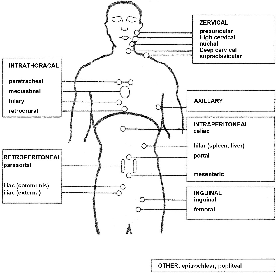
The Ann Arbor classification is used for staging [8] (Table 1). This requires anamnesis of B symptoms, a physical examination of the tonsils, lymph nodes, liver, spleen, effusions, visible or palpable masses, computed tomography (CT) with contrast medium of the neck, thorax, and abdomen, and a bone marrow biopsy (if no positron emission tomography (PET/CT) has been performed for staging; unilateral; aspiration and trephine of at least 2 cm in length). The definition of the lymph node regions is shown in Figure 1.
The most reliable method for detecting lymphoma manifestations is PET/CT using the tracer 18-fluorodeoxyglucose (FDG). FDG-PET is the international standard for staging and evaluating treatment outcomes [9]. The spread of the disease is usually diagnosed using whole-body PET/CT, in which pathological glucose accumulation (PET component) is assigned to anatomical structures (CT component). When a PET/CT is used, the results of the spread diagnosis are described using the Lugano classification based on the Ann Arbor classification [9]. PET/CT can provide additional information for diagnosis regarding bone marrow infiltration and staging and leads to so-called "upstaging" in approximately 20% of cases. The metabolically active volume at diagnosis is an additional prognostic parameter.
Stage | Definition |
|---|---|
I | Nodal involvement of a single lymph node region |
IE | Nodal involvement of a single lymph node region with growth into extranodal tissue (per continuitatem) or presence of a single primary extranodal focus |
II | Invasion of multiple lymph node regions on one side of the diaphragm |
IIE | Involvement of multiple lymph node regions on one side of the diaphragm with invasion into extranodal tissue (per continuitatem) |
III | Involvement of lymph node regions on both sides of the diaphragm |
IIIE | Involvement of lymph node regions on both sides of the diaphragm with invasion (per continuitatem) into extranodal tissue |
IV | Diffuse or disseminated involvement of one or more extralymphatic organs: |
Lymphatic tissue includes: lymph nodes, tonsils, Waldeyer's pharyngeal ring, Peyer's patches, and the spleen. | |
Suffix S | Involvement of the spleen (considered a lymph node) |
Addition A | None of the general symptoms defined under B |
Addition B | One or more of the following three general symptoms:
|
4.3Laboratory tests
Laboratory tests include a complete blood count with differential blood count and clinical chemistry tests to assess liver (bilirubin, GOT, GPT, alkaline phosphatase, gamma-GT) and kidney function (creatinine). Serum lactate dehydrogenase (LDH) provides information about cell proliferation and turnover, while uric acid provides information about cell breakdown. With regard to therapy-related complications, the heart should be examined (electrocardiogram, echocardiography). Furthermore, hepatitis and HIV serology should be performed in all patients before starting therapy.
4.4Prognostic factors
4.4.1International Prognostic Index (IPI)
The prognosis can be estimated using the International Prognostic Index (IPI), which takes into account the factors age (≤ vs. > 60 years), general condition (ECOG 0-1 vs. ≥ 2), Ann Arbor stage (I, II vs. III, IV), extranodal organ involvement (0-1 vs. ≥ 2 extranodal organs) and LDH (≤ vs. > upper normal limit) in favorable vs. unfavorable forms, with 0 vs. 1 point awarded in each case [10]. The calculated IPI score is subsequently used to distinguish between four risk groups: 0-1 points: low risk (overall survival after 3 years: 91%); 2 points: low-intermediate (81%); 3 points: high-intermediate (65%); 4-5 points: high (59%).
The age-adjusted International Prognostic Index (aaIPI) is a prognosis score based on the factors general condition, Ann Arbor stage, and LDH, in which the four risk groups mentioned above are defined by 0, 1, 2, or 3 unfavorable factor scores. In contrast to the IPI, the aaIPI allows for an age-independent survival prognosis.
A further development of the IPI is the National Comprehensive Cancer Network (NCCN) IPI, in which age and LDH levels are divided into 4 and 3 subgroups, respectively, and only a few extranodal manifestations are considered risk factors [11]. The NCCN-IPI separates the survival curves of the risk groups better than the original IPI. However, due to its complexity, it has not yet become widely accepted.
4.4.2Bone marrow involvement
According to a retrospective study, large cell (concordant) bone marrow infiltration is a risk factor independent of the International Prognostic Index [12]. This does not apply to small cell (discordant) infiltration, which may be an expression of an indolent lymphoma component. In cases of discordant bone marrow infiltration, relapses can manifest as aggressive or indolent lymphoma. The progression-free survival of patients with discordant infiltration is shorter than that of patients without bone marrow involvement, but the overall survival does not differ [12].
4.4.3Bulk
4.4.4Comorbidities
An assessment of comorbidities should always be performed before starting therapy. Both the Charlson Comorbidity Score and the Hematopoietic Cell Transplantation-specific Comorbidity Index (HCT-CI) are suitable for this purpose, although the HCT-CI is more accurate with regard to cardiovascular and nephrological comorbidities. Both scores are predictive and prognostic factors. In addition, if therapy is intensified at a later stage in the event of relapse or refractoriness, a comparison of comorbidities at diagnosis and at relapse can be helpful in making a treatment decision [15, 16].
4.5Differential diagnosis
Diffuse large B-cell lymphoma, with its variants and subtypes, and diseases related to diffuse large B-cell lymphoma must be distinguished from lymphomas that manifest similarly but are treated according to different principles. These include Hodgkin's lymphoma, Burkitt's lymphoma, mantle cell lymphoma, and peripheral T-cell lymphomas. The histological differentiation of diffuse large B-cell lymphoma from other aggressive B-cell lymphomas is based on the WHO classification [1].
5Therapy
5.1Treatment structure
Diffuse large B-cell lymphoma and related entities are generally curable diseases, but if left untreated, they can quickly become fatal. The indication for therapy is based on the diagnosis. Treatment is carried out with a curative intent. Exceptions are situations in which comorbidities or other circumstances do not allow for a curative approach. The following sections describe the current standard of care based on the latest results from clinical studies. First-line therapy continues to be based on the use of immunochemotherapy. Innovative immunotherapy concepts are used for the treatment of recurrence. The treatment structure is presented in separate algorithms for first-line treatment (Figure 2) and the various constellations in the case of relapse (Figure 3). To improve treatment, patients with diffuse large B-cell lymphoma should be treated in prospective studies whenever possible.
Since chemotherapy can lead to infertility, patients should be informed about fertility preservation measures before starting treatment and these should be initiated if necessary.
5.1.1First-line therapy
After completion of first-line therapy, radiotherapy should be considered for localized PET-positive residual manifestations.
The therapy protocols are described in the corresponding document "Systemic tumor therapy (German only)."
5.1.1.1Immunochemotherapy
First-line therapy consists of 6 cycles of the CHOP protocol and 8 doses of rituximab (R-CHOP protocol) or protocols similar to R-CHOP [17- 19]. The CHOP protocol is equivalent to more complex therapy regimens with better tolerability [20]. The addition of rituximab improved treatment outcomes in all subgroups studied [17- 21]. Randomized studies tested two variants of the R-CHOP protocol, which deliver comparable results in terms of progression-free survival and overall survival [17- 19]: 8 cycles at 21-day intervals or 6 cycles at 14-day intervals followed by 2 additional doses of rituximab. The latter variant requires the administration of G-CSF. Various analyses showed that 6 cycles at 21-day intervals are not inferior to 8 cycles of R-CHOP [22]. Six cycles of R-CHOP-21 are therefore the international standard. In younger patients (aged 60 years and younger) without IPI risk factors and without bulk, therapy should be reduced to 4 cycles of R-CHOP with 2 additional doses of rituximab without loss of efficacy [21]. In older patients (aged 61 years and older) without other risk factors (IPI 1, no bulk ≥7.5 cm), chemotherapy can also be discontinued after 4 cycles for patients in complete metabolic remission determined by PET-CT, but this should be followed by 4 additional doses of rituximab. Patients with PET-positive disease after 4 x R-CHOP should receive a total of 6 cycles of R-CHOP and 2 x rituximab followed by radiotherapy of the initial lesions [23]. In patients at increased risk (IPI 2-5), 6 doses of R-CHP in combination with polatuzumab vedotin followed by two doses of rituximab showed significantly improved progression-free survival compared to standard therapy with 6 doses of R-CHOP and two doses of rituximab (see also comments in chapter 5.1.1.4.3) [24]. Retrospective subgroup analyses show a preferential efficacy of 6 doses of R-CHP in combination with polatuzumab vedotin, especially in ABC DLBCL patients. However, since determination of the COO subtype by gene expression analysis is not currently standard practice in routine clinical care, these results have no immediate therapeutic consequences at present.
In addition to the therapies described, intensified protocols can also be used in younger high-risk patients, as described in chapter 5.1.1.4.3.
5.1.1.2Maintenance therapy
Maintenance therapy is not indicated in diffuse large B-cell lymphoma, as neither rituximab nor other substances (such as lenalidomide) have led to an improvement in overall survival.
5.1.1.3Radiation
The significance of radiation therapy in the treatment concept for diffuse large B-cell lymphoma patients has not been confirmed by randomized studies. Historically, radiation therapy has evolved from a sole curative treatment modality to a consolidating therapy in the sense of "involved field" radiation after completion of chemotherapy. Currently, the indication for consolidative radiotherapy is guided by a PET scan after completion of chemoimmunotherapy. Isolated PET-positive areas with a Deauville score of 4 or 5 should be treated with a total dose of 30 Gy in the form of radiation of the residual lymphoma ("residual mass" or "iceberg RT") [25]. The clinical target volume is defined as follows: residual lymphoma(s) plus a 1.5 cm safety margin.
In localized stages (I, II) of aggressive lymphomas, randomized studies showed that consolidation radiotherapy improved relapse-free survival but did not improve overall survival [26, 27]. In advanced stages, however, radiation of PET-positive residual tumors in initial bulk lesions (≥7.5 cm) led to a significant improvement in relapse-free survival and overall survival [14]. Retrospective studies suggest that this also applies to radiation of skeletal manifestations. At the end of chemotherapy, consolidative radiotherapy should be considered for individual PET-positive lesions.
The above-mentioned approach of consolidative radiotherapy for the treatment of PET-positive residual manifestations has been incorporated into numerous guidelines. With one exception, this approach is not covered by prospective randomized studies, but is based on retrospective analyses with historical controls [25, 28, 29]. General radiotherapy of initial lymphoma manifestations, as previously recommended as "involved field" radiotherapy, is obsolete [30].
5.1.1.4Unresolved questions in the field of first-line therapy
5.1.1.4.1Prognosis-driven therapy
The extent to which patients with aggressive lymphomas that differ in terms of gene expression or genetic subtype benefit from a change in therapy has not yet been conclusively clarified. The long-term results of the REMoDL-B study showed a significant improvement in progression-free and overall survival with the addition of the proteasome inhibitor bortezomib to R-CHOP in patients with diffuse large B-cell lymphoma of the ABC subtype [31]. In retrospective studies, more intensive treatment protocols (e.g., DA-EPOCH-R, Burkitt protocols) were beneficial in double-hit lymphomas. For example, DA-EPOCH-R prolonged progression-free and overall survival compared to R-CHOP in a non-randomized Canadian cohort study with 66 (treated 2015-2020) vs. 38 (treated 2005-2010) patients [32]. However, a clear treatment recommendation for intensified protocols cannot be derived from the currently available data for double-hit lymphomas.
5.1.1.4.2Response-guided therapy
It is currently unknown whether and under what conditions the results of interim staging should influence the further treatment strategy. Although the interim PET scan performed after 2 cycles of R-CHOP was of prognostic significance in a randomized study, intensification of treatment in the presence of unfavorable interim PET findings did not lead to an improvement in therapy compared to continuation of R-CHOP [20]. In contrast, a PET-guided French phase III study showed that early intensification in cases of slow response (PET still positive after two cycles, negative after four cycles) with high-dose therapy and autologous stem cell transplantation led to the same results as standard therapy completed in cases of early PET negativity [33].
5.1.1.4.3Complex therapy protocols
In patients below the age of 60 with an intermediate prognosis (aaIPI 1), the R-ACVBP protocol proved to be significantly superior to the R-CHOP protocol [34]. The therapeutic approach is similar to that commonly used for patients with acute lymphocytic leukemia, in that induction with 4 cycles of a dose-intensified R-CHOP variant is followed by consolidation with 2 cycles of high-dose methotrexate, 4 cycles of rituximab/ifosfamide/etoposide, and 2 cycles of cytarabine. In younger patients with an unfavorable prognosis (aaIPI 2 or 3), R-ACVBP was not superior to R-CHOP in a randomized, interim PET-guided study [35]. Due to increased toxicity in older patients, the R-ACVBP protocol has not become widely accepted. However, the protocol could offer advantages for patients at high risk of central nervous system relapse, as central nervous system relapses occur less frequently compared to R-CHOP [36].
In the randomized phase 3 POLARIX study, patients aged 18 to 80 years with intermediate and high risk (IPI 2-5) were randomized between 6 cycles of R-CHOP and 6 doses of R-CHP in combination with the anti-CD79B antibody-drug conjugate (ADC) polatuzumab vedotin, each followed by two applications of rituximab [24]. Patients treated with R-CHP-polatuzumab vedotin in the experimental arm showed a significant improvement in the primary endpoint of progression-free survival after 2 years (77% vs. 70%) [24]. To date, no difference in overall survival has been observed even after a longer observation period [37]. Furthermore, no increased toxicity was observed in the experimental arm. Polatuzumab vedotin in combination with R-CHP has been approved by the EMA as first-line therapy for the treatment of adults with diffuse large B-cell lymphoma.
The addition of etoposide (100 mg/m² days 1-3) to the CHOP protocol led to an improvement in event-free survival in younger patients with a good prognosis in the pre-rituximab era [38]. After the addition of rituximab, however, the results of CHOP and CHOEP were comparable [18]. In younger patients with a poor prognosis, unexpectedly good results were achieved with the R-CHOEP protocol. Patients treated with R-CHOEP achieved a 10-year overall survival rate of 72% [39]. These results suggest that patients with a poor prognosis could also benefit from the addition of etoposide in the rituximab era. A retrospective comparison of the results of younger patients with an unfavorable prognosis who were treated with either R-CHOEP or R-CHP in combination with polatuzumab vedotin showed no differences in progression-free and overall survival, but less acute toxicity with R-CHP in combination with polatuzumab vedotin [40]. Despite methodological limitations, these data support the cautious use of R-CHOEP in this population.
The complex B-ALL/NHL protocol of the German ALL Study Group includes, in addition to the alkylating agents, anthracyclines, vinca alkaloids, and corticosteroids contained in the CHOP protocol, the cerebrospinal fluid-penetrating substances methotrexate, cytarabine, and etoposide. Compared to the CHOP protocol, is characterized by higher toxicity (especially mucositis) with comparable therapy-associated mortality. In cases of poor response to the first two R-CHOP cycles, switching to the B-ALL/NHL protocol under randomized conditions showed no advantage over continuing with R-CHOP [20]. Therefore, there is no clear evidence to justify the use of the B-ALL/NHL protocol in patients with diffuse large B-cell lymphoma.
Given the importance of high-dose therapy with autologous blood stem cell transplantation in patients with recurrent lymphoma, numerous attempts have been made to use the procedure as consolidation therapy in primary treatment. Overall, no advantages could be demonstrated, and in some subgroups the results were contradictory [41]. Outside of clinical trials, consolidative high-dose therapy with autologous blood stem cell transplantation is not recommended as a concept for first-line therapy.
5.1.1.4.4Rituximab dosage and new CD20 antibodies
In a retrospective analysis, men benefited less than women from the addition of rituximab to the CHOP protocol. The reason for this appears to be faster rituximab clearance [42]. By increasing the single dose of rituximab from 375 mg/m² to 500 mg/m², progression-free survival in men was improved compared to a historical control [43]. In a second historical comparison, changing the timing of rituximab administration resulted in improved treatment outcomes in older patients [44]. These observations are contradicted by the results of prospective studies [20, 45] and retrospective comparisons, which showed no survival benefit from additional rituximab administration. There are no generally accepted recommendations for optimizing the use of rituximab.
New CD20 antibodies such as obinutuzumab [46] or ofatumumab [47] did not improve treatment outcomes compared to rituximab in randomized trials and are therefore not recommended.
5.1.1.4.5Vitamin D
Retrospective data suggested that patients with diffuse large B-cell lymphoma and serum vitamin D concentrations below the normal range would have a less favorable disease course than patients with normal vitamin D concentrations [48]. It is not known whether treatment outcomes can be improved by vitamin D supplementation, but laboratory-detected reduced vitamin D levels should be corrected.
5.1.2Therapy for progression/refractoriness/relapse
The description of the treatment protocols can be found in the corresponding document "Systemic tumor therapy."
5.1.2.1Therapy in the first relapse – early relapse and refractory situation
5.1.2.1.1CAR T-cell therapy
In recent decades, conventional platinum-based salvage therapy followed by high-dose therapy with autologous blood stem cell transplantation has been the standard therapy for relapse in younger patients (under 60 years of age) as well as in older patients without therapy-limiting comorbidities [49]. Satisfactory treatment results were generally only achieved if the relapse responded to conventionally dosed induction therapy. With an interval of less than 12 months between primary diagnosis and relapse, this was rarely the case. Three cycles of the R-DHAP or R-ICE protocol proved to be equivalent as induction therapy [50]. Alternatively, the R-GDP regimen can be used, which is equivalent to the R-DHAP protocol but is better tolerated [51]. The BEAM protocol was generally used for high-dose therapy [50].
For younger patients eligible for high-dose therapy with primarily refractory disease or early relapse (within 12 months after completion of first-line therapy), the above-mentioned standard (high-dose therapy followed by autologous stem cell transplantation) was directly compared with anti-CD19 CAR T-cell therapy in various randomized phase 3 studies [52- 54]. The ZUMA-7 study showed a significant improvement in event-free and overall survival for the experimental arm with axicabtagen ciloleucel in patients with diffuse large B-cell lymphoma [52]. In the TRANSFORM study, the experimental arm with lisocabtagene maraleucel also led to a significant improvement in event-free survival and to the approval of lisocabtagene maraleucel in patients with diffuse large B-cell lymphoma, high-grade B-cell lymphoma (HGBCL), primary mediastinal B-cell lymphoma, and follicular large B-cell lymphoma [53]. In this respect, therapy with axicabtagen ciloleucel or lisocabtagen maraleucel represents the new standard in patients with primary refractory disease or early relapse. In the approval studies as well as in real-world analyses, lisocabtagene maraleucel showed a better toxicity profile in indirect comparison to axicabtagene ciloleucel [55]. For information on the management of CAR T-cell-associated toxicities, please refer to Onkopedia CAR T-cells: Management of side effects (German only).
Non-relapse mortality after CAR T-cell therapies is approximately 5% in long-term observation and is often caused by cytopenia and immunodeficiency-induced infections or secondary neoplasms. Therefore, follow-up care should either be carried out directly at the CAR T-cell centers or closely coordinated with the responsible center.
CAR T-cell therapy for primarily refractory disease or early relapse should also be offered to older patients who may not be eligible for high-dose therapy, as older patients can benefit from CAR T-cell therapy to a similar extent as younger patients. It should be noted that the inability to undergo CAR T-cell therapy is not uniformly defined internationally. The ALYCANTE study in patients who are not eligible for high-dose therapy showed promising results with axicabtagen ciloleucel in terms of metabolic response and progression-free survival. The use of lisocabtagene maraleucel in the PILOT study also showed favorable response and survival data in this patient population [56, 57].
5.1.2.1.2Non-CAR T-cell therapy approaches
For patients who are not eligible for high-dose therapy and are experiencing their first relapse, data from the STARGLO Phase III study now provide the first evidence on the combination of the bispecific antibody Glofitamab with chemotherapy. In the study, patients were randomized 2:1 between Glofitamab-Gemcitabine-Oxaliplatin (GemOx) and R-GemOx [58]. The combination of Glofitamab and GemOx significantly increased the complete remission rate (59% vs. 25%) and led to a significant improvement in overall survival (2-year OS: 54 months vs. 34 months) and 18-month PFS with good overall tolerability [58]. For patients in the second line, the survival benefits were even more pronounced.
Therefore, for patients who are not eligible for high-dose therapy in first relapse, the Glofitamab-GemOx regimen is an effective alternative therapy to CAR-T cell administration. However, unlike the ALYCANTE and PILOT studies, the STARGLO study did not include patients with HGBCL or transformed lymphomas, and approval is therefore limited to DLBCL-NOS.
Another phase III study (POLARGO) in patients not eligible for high-dose therapy in first relapse also showed a significant improvement in overall survival (44 months vs. 33 months) with the addition of polatuzumab vedotin to R-GemOx, but increased toxicity was observed in the experimental arm, so polatuzumab vedotin plus R-Gem-Ox will only be used for selected patients [59]. This is even more true in the primarily refractory or early recurrence situation, as patients should preferentially receive CAR-T cells or (if not eligible for high-dose therapy) Glofitamab-GemOx after first-line therapy containing polatuzumab.
The SUMNO study is the third phase III study in the segment of non-high-dose-eligible patients in first relapse, in which patients were randomized 2:1 to receive the mosunetuzumab-polatuzumab vedotin combination versus an R-GemOx comparator arm. With overall survival data still immature, the 12-month PFS as the primary endpoint for mosunetuzumab polatuzumab vedotin is 49% (R-GemOx 18%) and the complete remission rate is 51% (24% with R-GemOx) [60].
Overall, it should be noted critically in the three Phase III studies discussed here that the criteria for classification as "not eligible for high-dose therapy" were not uniformly defined.
5.1.2.2Treatment in first relapse – late relapse
For patients with late recurrence (at least 12 months after completion of first-line therapy) who are eligible for autologous blood stem cell transplantation, high-dose chemotherapy followed by autologous stem cell transplantation remains the standard of care.
In patients with late recurrence who are not eligible for high-dose therapy with autologous blood stem cell transplantation due to their age or comorbidities, or in patients with early recurrence who do not qualify for CAR T-cell therapy, the treatment goal is often palliative. A curative treatment concept appears possible if the interval between the primary diagnosis and the relapse is long and the disease responds to renewed therapy. In addition to more intensive chemotherapy salvage regimens such as R-GDP, R-DHAP, or R-ICE [50] prior to mostly BEAM-based high-dose therapy, the STARGLO study now shows that patients who are not eligible for high-dose therapy can be offered treatment with Glofitamab-GemOx with good results [58]. The extent to which the above-mentioned standard of high-dose therapy with autologous blood stem cell transplantation in late relapse of bispecific antibody-based second-line therapy could be called into question, also with regard to a possible curative potential, is currently unclear. Furthermore, the combination of rituximab, bendamustine, and the antibody-drug conjugate polatuzumab vedotin (Pola-BR) is approved for patients with at least one prior line of therapy. The approval study for Pola-BR showed a significant improvement in response rates, progression-free survival, and overall survival compared to rituximab and bendamustine [61]. Another option that has been approved is a completely chemotherapy-free treatment consisting of the anti-CD19 antibody tafasitamab in combination with lenalidomide for patients who are not eligible for high-dose therapy with at least one prior line of therapy [62]. Promising response and long-term data were obtained in the L-MIND study. Due to the large number of newly approved treatment modalities, attention to sequence is becoming increasingly important. When using tafasitamab, the effectiveness of downstream CD19-targeted CAR T-cell therapy is currently unclear. Furthermore, the quality of lymphocyte apheresis after bendamustine is significantly limited in retrospective studies [63].
5.1.2.3Therapy from the second relapse onwards
The EMA (European Medicines Agency) has currently approved three CAR T-cell products, axicabtagene ciloleucel, tisagenlecleucel, and lisocabtagene maraleucel, for patients who have undergone at least two prior therapies [64- 66]. According to the approval studies, the indication is for patients with relapsed/refractory diffuse large B-cell lymphoma, primary mediastinal B-cell lymphoma, or transformed follicular lymphoma. In patients with a second relapse, the possibility of CAR T-cell treatment should therefore always be considered.
CAR T-cell therapy should also be offered to older patients, including those with comorbidities. The suitability of CAR T-cell therapy should be evaluated on a case-by-case basis and product-specific. Real-world analyses for the use of all three approved CAR T-cell products in the third and higher lines of therapy show no negative influence of age on the success of CAR T-cell therapy, so that no upper age limit for the use of CAR T-cell therapy can generally be specified [67].
With regard to comorbidities, significant negative effects of moderate to severe renal, cardiac, and hepatic comorbidities have been described for axicabtagene ciloleucel, which must be taken into account when determining the indication. However, since successful use in patients with corresponding pre-existing conditions has been reported in smaller case series and prospective studies, and since there are usually no proven less toxic treatment modalities with curative potential, there is no absolute contraindication to the use of axicabtagene ciloleucel.
Comparisons between studies and existing real-world analyses show that tisagenlecleucel and lisocabtagen-maraleucel have significantly lower overall toxicity compared to axicabtagen-ciloleucel, resulting in low incidences of non-lymphoma-related mortality and the need for intensive care treatment [68]. For tisagenlecleucel, an analysis of 1,159 patients found no negative association between comorbidities and survival data [69]. The use of these two CAR T-cell products in their respective approved areas is therefore recommended even in the presence of relevant comorbidities due to the lack of less toxic alternatives with proven curative potential.
From the second relapse onwards, the bispecific antibodies epcoritamab, glofitamab, and odronextamab are available. Normally, they should be used after CAR T-cell therapy has failed. Bispecific antibodies induce response rates of approximately 50-60% from the second relapse onwards, with patients who achieve complete remission in particular remaining disease-free in the long term [70- 72]. The three approved bispecific antibodies differ in terms of their application, duration of therapy, and structure. Epcoritamab is administered subcutaneously for an unlimited period of time until progression or until the occurrence of intolerable side effects, Odronextamab is administered intravenously also until progression or until the occurrence of intolerable side effects, while Glofitamab is administered intravenously for a limited period of 12 cycles. The curative potential of epcoritamab, glofitamab, and odronextamab cannot yet be conclusively determined. The above-mentioned Glofitamab-GemOx regimen is also available for this indication and produces plateau-like remissions with short follow-up. The significance of adding GemOx-based chemotherapy to Glofitamab monotherapy from the second relapse onwards is not yet clear [58].
Another therapeutic option for patients after failure of CAR T-cell therapy may be allogeneic stem cell transplantation in suitable patients, depending on the remission status prior to stem cell transplantation. If complete metabolic remission cannot be achieved after failure of CAR T-cell therapy, e.g., with a bispecific antibody, allogeneic stem cell transplantation may be considered in individual cases for fit patients [73].
Furthermore, the anti-CD19 antibody-drug conjugate loncastuximab tesirin is available for treatment from the second relapse onwards [74]. Approximately 50% of patients with previously treated diffuse large B-cell lymphoma respond to treatment with loncastuximab tesirin.
The ViPOR regimen (combination of venetoclax, ibrutinib, obinutuzumab, lenalidomide, and prednisolone) represents an unapproved treatment option for patients with chemotherapy-refractory or multiple relapsed disease [75]. In a phase 1b/2 study on the tolerability and efficacy of ViPOR, an overall response rate of 54% was observed; in line with the molecular vulnerabilities addressed, patients with non-GCB subtype and high-grade B-cell lymphoma with MYC and BCL2 or BCL6 translocation benefit most.
5.1.3Significance of salvage and bridging therapy prior to CAR T-cell administration
Many patients with progressive or relapsed disease are in a clinically reduced state. Already in the CAR T-cell approval studies from the third line onwards, but also in the second-line situation, preparatory therapies were used in some patients to enable a response and thus also improve the clinical condition of the patients. For lisocabtagen maraleucel and tisagenlecleucel, bridging concepts were part of the approval studies, whereas the use of bridging therapy was excluded from the studies using axicabtagen ciloleucel. There are no data from prospective comparative studies on the necessity of bridging therapy or on the question of the necessary intensity or the treatment modalities and substances to be used. Retrospective studies have shown that approximately 80% of all patients receive bridging or salvage therapy prior to CAR T-cell therapy. The prognosis is significantly better in patients who respond to bridging therapy compared to those who do not respond [76, 77]. However, retrospective analyses have found no evidence that particularly intensive immunochemotherapy is beneficial as bridging therapy [78].
With the above limitations in mind, the following recommendations can be made:
Salvage/bridging therapy should be used if patients are at risk of becoming clinically unstable during the preparatory period.
Higher-grade organ toxicities caused by intensive chemotherapy are probably not justified and should be avoided.
Studies investigating the value of various substances as components of bridging therapy have provided evidence of a beneficial effect of polatuzumab vedotin as a component of bridging therapy [79]. It is a valid option, especially for patients who have not already received polatuzumab vedotin.
Bendamustine has a quantitatively and qualitatively unfavorable effect on the quality of leukocyte apheresis, which negatively impacts the overall success of the therapy [63, 80]. Therefore, the use of bendamustine should be avoided prior to apheresis.
Despite potentially negative aspects, bispecific antibodies can be used prior to CAR T-cell therapy [81]. However, there is no evidence of a specifically beneficial effect to date. Its use should be considered in particular in cases of progression following immediately prior immunochemotherapy.
Radiotherapy can be used as bridging therapy, especially in cases of limited lymph node involvement. This can be particularly useful if no previous radiotherapy has been administered and due to the often favorable toxicity profile and, in some cases, improved antigen presentation [82]. The usual radiation dose is 30 Gy in 15 fractions. In cases of very large lymphoma manifestations (>10 cm), 40 Gy can be administered. If the timeline does not allow for three weeks of therapy, 20 Gy in 5 fractions is also possible.
5.2Special lymphomas and clinical situations
5.2.1Primary mediastinal B-cell lymphoma (PMBL)
5.2.1.1General
Primary mediastinal B-cell lymphoma (PMBL) accounts for approximately 2-4% of all lymphomas and is classified as an independent entity by the WHO due to its different clinical and pathological characteristics [1]. In terms of molecular pathogenesis, PMBL shows similarities to classic Hodgkin's lymphoma (e.g., frequent CD30 positivity, constitutive activation of the NF-κB and JAK/STAT signaling pathways, and PD-1-mediated immune evasion). PMBLs occur primarily in young women (median age approximately 35 years). Patients with PMBL often present with symptoms of upper respiratory congestion or airway compression due to the extent or locally invasive growth of the mediastinal mass.
5.2.1.2Primary therapy
Patients with PMBL are often treated with the same protocols as patients with diffuse large B-cell lymphoma. These protocols achieve very high cure rates [83, 84]. Unlike diffuse large B-cell lymphoma, however, the optimal chemotherapy regimen for first-line treatment of PMBL has not yet been definitively established. Accordingly, there are currently two predominant options: R-CHOP and DA-EPOCH-R. The DA-EPOCH-R protocol, which contains the same substances as the R-CHOEP protocol but differs in the method of administration (96-hour continuous infusion of etoposide, vincristine, and doxorubicin) and an adjustment of the cytostatic doses to the granulocyte and thrombocyte nadir of the previous cycle, achieved an overall survival rate of 97% (95% CI 81-99) after 5 years in a single-arm study [84]. PMBL patients treated in real-world analyses often received consolidating involved field radiation of residual tumor after first-line therapy. A PET-CT scan should be performed to clarify whether radiation is necessary after systemic therapy. The IELSG 37 study showed that if PET-CT is negative after systemic therapy, radiation can be omitted [85].
5.2.1.3 Recurrence therapy
The high effectiveness of first-line PMBL therapy results in a relatively low proportion of primarily refractory or relapsed cases, which makes it difficult to systematically develop an optimal therapy for relapse or refractoriness. Patients with primarily refractory disease or early relapse (within 12 months after completion of first-line therapy) should undergo CAR T-cell therapy with lisocabtagen maraleucel [53]. Patients who experience late relapse (more than 12 months after completion of first-line therapy) after an initial chemosensitive disease should be treated with platinum-based salvage therapy (R-DHAP, R-GDP, or R-ICE) and, upon achieving at least partial remission, with high-dose therapy according to the BEAM protocol followed by autologous stem cell transplantation. In a primarily chemo-refractory situation, treatment with PD-1 blockade with or without brentuximab vedotin is an option [86- 88]. However, PD-1 blockade is only approved for this indication in the US and Switzerland; in Germany, it is used off-label. Anti-CD19 CAR T-cell therapy remains an option for the third line of therapy if CAR T-cell therapy has not yet been used in the second line of therapy.
5.2.2Richter transformation (RT)
Richter transformation (also known as Richter syndrome) refers to the development of aggressive lymphoma from chronic lymphocytic leukemia (CLL), which can occur in 2-10% of CLL cases during the course of the disease. Clinically, these patients often show rapid deterioration with pronounced B symptoms and massive lymphadenopathy, accompanied by an increased LDH. If Richter transformation is suspected, patients should undergo FDG-PET/CT to enable rapid excision of a metabolically highly active lymph node. Well over 90% of cases show diffuse large B-cell lymphoma histology, while the remaining cases are mostly histologically diagnosed as Hodgkin's lymphoma. If possible, to differentiate between RT and de novo secondary lymphoma, it should be investigated whether there is a clonal relationship. The standard therapy for RT with diffuse large B-cell lymphoma histology is 4 to 6 cycles of R-CHOP, with an overall response rate of only about 40% and a very short median survival of about 6-8 months [89]. For younger patients with RT, a consolidating allogeneic stem cell transplant is usually recommended. For the few cases with RT and Hodgkin histology, therapy based on standard Hodgkin protocols is recommended, even though data from larger treatment series are lacking.
In small case series, a potential therapeutic benefit of BTK inhibitors such as acalabrutinib and pirtobrutinib in RT has been demonstrated, although only very short progression-free survival of a few months have been reported. Checkpoint inhibitors, in particular pembrolizumab, also showed responses as monotherapy in RT. These individual therapy options (all off-label) can be considered in cases of limited fitness or after failure of R-CHOP-based therapies.
In a phase II study by the DCLLSG (RT-1 study), a response rate of 58% of treated patients was documented using the combination of the PD-1 inhibitor tislelizumab with the second-generation BTK inhibitor zanubrutinib, with a median progression-free survival of 10 months and approximately 75% of patients still alive after one year [90]. This unapproved combination therapy is a promising alternative, especially for older patients who are not eligible for stem cell transplantation.
Furthermore, the use of CAR T cells in RT is currently being investigated. Initial results based on axicabtagene ciloleucel, tisagenlecleucel, lisocabtagene maraleucel, and brexucabtagene autoleucel indicate high response rates in small case series (ORR 63%; CR rate 46%), but with little reliable long-term results (2-year PFS and OS of 33% and 47%, respectively) [91, 92].
5.2.3First-line therapy in the elderly
5.2.4Central nervous system manifestations
Following first-line therapy with R-CHOP, approximately 2-5% of patients with diffuse large B-cell lymphoma experience a relapse of the disease in the central nervous system (CNS).
Since central nervous system relapses are rare and the evidence is inconclusive, general CNS prophylaxis is not recommended. The CNS-IPI, which consists of the 5 factors of the IPI and involvement of the kidneys and/or adrenal glands, is suitable for identifying patients at high risk of central nervous system relapse [94]. With 0-1 risk factors, the risk of CNS relapse is <1%; with 2-3 risk factors, it is 3%; and with 4-6 risk factors, it is 10%. In high-risk groups, targeted diagnostics (magnetic resonance imaging of the central nervous system, FACS analysis of cerebrospinal fluid) are recommended. The integration of molecular markers into the CNS-IPI showed that patients with a high CNS-IPI and an ABC or unclassifiable molecular subtype have a risk of central nervous system relapse of more than 15% [95]. There is currently no clear evidence for drug-based CNS prophylaxis, e.g., with high-dose methotrexate. Various retrospective analyses showed no advantage of CNS prophylaxis with high-dose methotrexate for the prevention of CNS relapse [96]. Intrathecal prophylaxis is not indicated.
Patients with parallel systemic and central nervous system involvement at diagnosis should be treated with therapy protocols that target both the peripheral and central nervous system lymphoma components. Protocols involving various substances and varying intensities are used here. If possible, autologous stem cell transplantation should be performed [97]. As with primary CNS lymphomas, CNS-permeable substances should be used.
5.2.5Testicular lymphoma
Testicular lymphoma is characterized by a high rate of relapse in the central nervous system and contralateral testicles. Based on retrospective data, CNS prophylaxis with high-dose methotrexate and irradiation of the contralateral testicle with 30 Gray are recommended in addition to standard therapy [98].
5.2.6Contraindication to anthracyclines
In cases of advanced heart failure or extensive prior anthracycline treatment, doxorubicin contained in the R-CHOP protocol cannot be used without significant risks. The pegylated liposomal doxorubicin formulation is considered to be equipotent but less cardiotoxic. Under liposomal doxorubicin, sensitive heart failure markers (ejection fraction, NT-proBNP) reach pathological values less frequently compared to non-pegylated doxorubicin. However, clinically manifest heart failure develops rarely under both doxorubicin preparations and with comparable frequency [99]. Etoposide (50 mg/m² IV on day 1, 100 mg/m² PO on days 2 and 3) offers an anthracycline-free alternative to doxorubicin. As an indication of its curative potential, a plateau in the survival curve is observed following R-CEOP, similar to that after R-CHOP. R-GCVP (gemcitabine instead of doxorubicin) has been studied in a population with cardiac morbidity, including heart failure and coronary heart disease: in older patients, it resulted in a response rate of 61% and a 2-year progression-free survival rate of 50% [100].
5.2.7Impaired renal or hepatic function
Since the active metabolites of the substances contained in the R-CHOP protocol are predominantly not eliminated renally, the protocol can generally also be applied in cases of impaired renal function. In cases of impaired liver function, the hepatically eliminated substances doxorubicin and vincristine accumulate. They should therefore not be used in cases of advanced liver dysfunction or bile duct obstruction. If the dysfunction is caused by lymphoma, it can often be reversed by one or two cycles of doxorubicin- and vincristine-free chemoimmunotherapy (e.g., rituximab 375 mg/m² on day 1; cyclophosphamide 200 mg/m² days 1-5; etoposide 100 mg/m² days 1-3; prednisone 100 mg days 1-5; modification of cyclophosphamide and etoposide treatment according to clinical condition).
5.2.8Pregnancy and fertility
If aggressive lymphoma occurs in the first trimester, termination of pregnancy is recommended, as chemotherapy during the organogenesis phase carries a high risk of malformations. The risk is low in the second and third trimesters. The R-CHOP protocol is suitable as a standard treatment regimen. Antimetabolites (e.g., methotrexate) must not be used due to the risk of fetal CNS damage. If lymphoma occurs in late pregnancy and is not very aggressive, treatment can be postponed until after birth.
A retrospective study showed no disadvantage for the mother if treatment of the lymphoma was not started until after delivery, and only a low risk for the fetus if therapy was carried out in the second or third trimester (rate of stillbirths and malformations approximately 5%) [101]. Prematurity impairs the cognitive development of the child. The indication for premature delivery should therefore be strictly defined.
Infertility is primarily caused by alkylating agents and radiation in the pelvic area. In some men treated with the CHOP protocol, spermatogenesis recovers within 5 to 7 years [102]. Since this cannot be predicted in individual cases, sperm should be preserved before chemotherapy if the patient wishes to undergo genetic counseling. In women, permanent amenorrhea rarely occurs following treatment with CHOP [103]. However, a reduction in ovarian reserve with premature menopause (last menstrual cycle before the age of 40) is common. The window of opportunity for fulfilling the desire to have children is particularly short for patients who are over the age of 30 at the time of chemotherapy [103].
6Follow-up and aftercare
6.1Follow-up
6.1.1Interim examination
During first-line or recurrence therapy, an interim examination is usually performed to confirm a potentially successful treatment strategy. The timing and method of the interim assessment and the extent of tumor reduction required for continuation of therapy have not been defined [9]. Interim staging is usually performed after one-third or one-half of the treatment protocol using computed tomography. Interim PET/CT examinations are a prognostic parameter in cases of persistent positivity [20]. Currently, the value of PET/CT-triggered therapeutic intensification has not been shown.
6.1.2Final examination
According to international standards, the treatment outcome is evaluated 6 to 8 weeks after the end of treatment using PET/CT, which is used to determine the response categories of complete remission (no FDG accumulation), partial remission (residual FDG accumulation with a reduction in tumor mass of at least 50%), stable disease (residual FDG accumulation with less than 50% reduction in tumor mass), and progressive disease (residual FDG accumulation with increase in the mass of one or more lymphoma manifestations or new FDG-positive lesion(s)) [9]. The Deauville classification is used to quantify the metabolic response.
If positron emission tomography is not used, the treatment outcome is defined by computed tomography based on the size of residual space-occupying lesions. Complete remission requires the absence of any residual space-occupying lesions. If the CT-based remission assessment alone is unclear, an additional PET/CT scan is recommended. Details of the response assessment, which can be difficult in individual cases, can be found in the original literature [9].
6.2Follow-up
Follow-up care serves to support reintegration into family, work, and society, to detect disease recurrence, and to identify and minimize long-term complications, particularly infertility, secondary malignancies, and cardiovascular disorders. According to the modified Cotswolds recommendations, follow-up care is provided at quarterly intervals for the first two years after the end of therapy, at six-monthly intervals for the following three years, and at annual intervals from the sixth year onwards. Follow-up examinations focus on medical history, physical examination, and laboratory analyses. Routine computed tomography or PET/CT scans are not recommended [9]. Imaging procedures are justified if the clinical findings suggest a relapse of the disease or a late complication.
In patients under the age of 45, the risk of developing a secondary malignancies is increased following successful treatment with CHOP-like therapy protocols [104]. In addition to myelodysplasia and acute myeloid leukemia, there are increased incidences of bronchial carcinoma (increased risk due to simultaneous nicotine consumption), colorectal carcinoma, prostate carcinoma, cyclophosphamide-induced bladder carcinoma, and Hodgkin's lymphoma. Specific measures for the prevention and detection of secondary malignancies have not been formulated for patients with diffuse large B-cell lymphoma.
After treatment with anthracyclines, the risk of developing heart failure is increased compared to the normal population, especially if chemotherapy was performed before the age of 55. Modifiable cofactors include arterial hypertension and nicotine consumption. If mediastinal or neck radiation is performed in addition to chemotherapy, the risk of coronary heart disease or cerebrovascular events also increases. To this end, it is recommended that cardiovascular risk factors (hypertension, hypercholesterolemia, diabetes mellitus, obesity, nicotine abuse) are taken care of. If young patients undergo mediastinal radiation, early breast cancer screening should be performed.
6.3COVID-19
Information on COVID-19 can be found in the Onkopedia COVID-19 guideline.
7[Kapitel nicht relevant]
8[Kapitel nicht relevant]
9References
Alaggio, Amador, Anagnostopoulos, et al., The 5th edition of the World Health Organization Classification of Haematolymphoid Tumours: Lymphoid Neoplasms. Leukemia, 2022. 36(7): p. 1720-1748. DOI:10.1038/s41375-022-01620-2
Sehn, Salles, Diffuse Large B-Cell Lymphoma. N Engl J Med, 2021. 384(9): p. 842-858. DOI:10.1056/NEJMra2027612
Morton, Wang, Devesa, et al., Lymphoma incidence patterns by WHO subtype in the United States, 1992-2001. Blood, 2006. 107(1): p. 265-76. DOI:10.1182/blood-2005-06-2508
Alizadeh, Eisen, Davis, et al., Distinct types of diffuse large B-cell lymphoma identified by gene expression profiling. Nature, 2000. 403(6769): p. 503-11. DOI:10.1038/35000501
de Jong, Rosenwald, Chhanabhai, et al., Immunohistochemical prognostic markers in diffuse large B-cell lymphoma: validation of tissue microarray as a prerequisite for broad clinical applications--a study from the Lunenburg Lymphoma Biomarker Consortium. J Clin Oncol, 2007. 25(7): p. 805-12. DOI:10.1200/jco.2006.09.4490
Rosenwald, Bens, Advani, et al., Prognostic Significance of MYC Rearrangement and Translocation Partner in Diffuse Large B-Cell Lymphoma: A Study by the Lunenburg Lymphoma Biomarker Consortium. J Clin Oncol, 2019. 37(35): p. 3359-3368. DOI:10.1200/jco.19.00743
Johnson, Slack, Savage, et al., Concurrent expression of MYC and BCL2 in diffuse large B-cell lymphoma treated with rituximab plus cyclophosphamide, doxorubicin, vincristine, and prednisone. J Clin Oncol, 2012. 30(28): p. 3452-9. DOI:10.1200/jco.2011.41.0985
Lister, Crowther, Sutcliffe, et al., Report of a committee convened to discuss the evaluation and staging of patients with Hodgkin's disease: Cotswolds meeting. J Clin Oncol, 1989. 7(11): p. 1630-6. DOI:10.1200/jco.1989.7.11.1630
Cheson, Fisher, Barrington, et al., Recommendations for initial evaluation, staging, and response assessment of Hodgkin and non-Hodgkin lymphoma: the Lugano classification. J Clin Oncol, 2014. 32(27): p. 3059-68. DOI:10.1200/jco.2013.54.8800
A predictive model for aggressive non-Hodgkin's lymphoma. N Engl J Med, 1993. 329(14): p. 987-94. DOI:10.1056/nejm199309303291402
Zhou, Sehn, Rademaker, et al., An enhanced International Prognostic Index (NCCN-IPI) for patients with diffuse large B-cell lymphoma treated in the rituximab era. Blood, 2014. 123(6): p. 837-42. DOI:10.1182/blood-2013-09-524108
Sehn, Scott, Chhanabhai, et al., Impact of concordant and discordant bone marrow involvement on outcome in diffuse large B-cell lymphoma treated with R-CHOP. J Clin Oncol, 2011. 29(11): p. 1452-7. DOI:10.1200/jco.2010.33.3419
Pfreundschuh, Ho, Cavallin-Stahl, et al., Prognostic significance of maximum tumor (bulk) diameter in young patients with good-prognosis diffuse large-B-cell lymphoma treated with CHOP-like chemotherapy with or without rituximab: an exploratory analysis of the MabThera International Trial Group (MInT) study. Lancet Oncol, 2008. 9(5): p. 435-44. DOI:10.1016/s1470-2045(08)70078-0
Held, Murawski, Ziepert, et al., Role of radiotherapy to bulky disease in elderly patients with aggressive B-cell lymphoma. J Clin Oncol, 2014. 32(11): p. 1112-8. DOI:10.1200/jco.2013.51.4505
Charlson, Pompei, Ales, et al., A new method of classifying prognostic comorbidity in longitudinal studies: development and validation. J Chronic Dis, 1987. 40(5): p. 373-83. DOI:10.1016/0021-9681(87)90171-8
Artz, Pollyea, Kocherginsky, et al., Performance status and comorbidity predict transplant-related mortality after allogeneic hematopoietic cell transplantation. Biol Blood Marrow Transplant, 2006. 12(9): p. 954-64. DOI:10.1016/j.bbmt.2006.05.015
Coiffier, Thieblemont, Van Den Neste, et al., Long-term outcome of patients in the LNH-98.5 trial, the first randomized study comparing rituximab-CHOP to standard CHOP chemotherapy in DLBCL patients: a study by the Groupe d'Etudes des Lymphomes de l'Adulte. Blood, 2010. 116(12): p. 2040-5. DOI:10.1182/blood-2010-03-276246
Pfreundschuh, Trümper, Osterborg, et al., CHOP-like chemotherapy plus rituximab versus CHOP-like chemotherapy alone in young patients with good-prognosis diffuse large-B-cell lymphoma: a randomized controlled trial by the MabThera International Trial (MInT) Group. Lancet Oncol, 2006. 7(5): p. 379-91. DOI:10.1016/s1470-2045(06)70664-7
Pfreundschuh, Schubert, Ziepert, et al., Six versus eight cycles of bi-weekly CHOP-14 with or without rituximab in elderly patients with aggressive CD20+ B-cell lymphomas: a randomized controlled trial (RICOVER-60). Lancet Oncol, 2008. 9(2): p. 105-16. DOI:10.1016/s1470-2045(08)70002-0
Dührsen, Müller, Hertenstein, et al., Positron Emission Tomography-Guided Therapy of Aggressive Non-Hodgkin Lymphomas (PETAL): A Multicenter, Randomized Phase III Trial. J Clin Oncol, 2018. 36(20): p. 2024-2034. DOI:10.1200/jco.2017.76.8093
Poeschel, Held, Ziepert, et al., Four versus six cycles of CHOP chemotherapy in combination with six applications of rituximab in patients with aggressive B-cell lymphoma with favorable prognosis (FLYER): a randomized, phase 3, non-inferiority trial. Lancet, 2019. 394(10216): p. 2271-2281. DOI:10.1016/s0140-6736(19)33008-9
Sehn, Congiu, Culligan, et al., No Added Benefit of Eight Versus Six Cycles of CHOP When Combined with Rituximab in Previously Untreated Diffuse Large B-Cell Lymphoma Patients: Results from the International Phase III GOYA Study. Blood, 2018. 132(Supplement 1): p. 783-783. DOI:10.1182/blood-2018-99-116845
Viola Pöschel, Excellent outcome with interim PET-adapted treatment reduction in elderly DLBCL patients with favorable prognosis: results of 288 patients treated in the OPTIMAL>60 trial of the DSHNHL/GLA. EHA2025, 2025.
Tilly, Morschhauser, Sehn, et al., Polatuzumab Vedotin in Previously Untreated Diffuse Large B-Cell Lymphoma. N Engl J Med, 2022. 386(4): p. 351-363. DOI:10.1056/NEJMoa2115304
Freeman, Savage, Villa, et al., Long-term results of PET-guided radiation in patients with advanced-stage diffuse large B-cell lymphoma treated with R-CHOP. Blood, 2021. 137(7): p. 929-938. DOI:10.1182/blood.2020005846
Stephens, Li, LeBlanc, et al., Continued Risk of Relapse Independent of Treatment Modality in Limited-Stage Diffuse Large B-Cell Lymphoma: Final and Long-Term Analysis of Southwest Oncology Group Study S8736. J Clin Oncol, 2016. 34(25): p. 2997-3004. DOI:10.1200/jco.2015.65.4582
Held, Zeynalova, Murawski, et al., Impact of rituximab and radiotherapy on outcome of patients with aggressive B-cell lymphoma and skeletal involvement. J Clin Oncol, 2013. 31(32): p. 4115-22. DOI:10.1200/jco.2012.48.0467
Persky, Unger, Spier, et al., Phase II study of rituximab plus three cycles of CHOP and involved-field radiotherapy for patients with limited-stage aggressive B-cell lymphoma: Southwest Oncology Group study 0014. J Clin Oncol, 2008. 26(14): p. 2258-63. DOI:10.1200/jco.2007.13.6929
Persky, Li, Stephens, et al., Positron Emission Tomography-Directed Therapy for Patients With Limited-Stage Diffuse Large B-Cell Lymphoma: Results of Intergroup National Clinical Trials Network Study S1001. J Clin Oncol, 2020. 38(26): p. 3003-3011. DOI:10.1200/jco.20.00999
Berger, Trelle, Büchi, et al., Impact on survival through consolidation radiotherapy for diffuse large B-cell lymphoma: a comprehensive meta-analysis. Haematologica, 2021. 106(7): p. 1923-1931. DOI:10.3324/haematol.2020.249680
Davies, Barrans, Stanton, et al., Differential Efficacy From the Addition of Bortezomib to R-CHOP in Diffuse Large B-Cell Lymphoma According to the Molecular Subgroup in the REMoDL-B Study With a 5-Year Follow-Up. J Clin Oncol, 2023. 41(15): p. 2718-2723. DOI:10.1200/jco.23.00033
Alduaij, Sehn, Champagne, et al., 75 | Population-wide introduction of dose-adjusted Epoch-R in high-grade B-Cell Lymphoma with MYC and BCL2 rearrangements with DLBCL morphology. Hematological Oncology, 2025. 43(S3): p. e75_70093. DOI:10.1002/hon.70093_75
Le Gouill, Ghesquières, Oberic, et al., Obinutuzumab vs rituximab for advanced DLBCL: a PET-guided and randomized phase 3 study by LYSA. Blood, 2021. 137(17): p. 2307-2320. DOI:10.1182/blood.2020008750
Récher, Coiffier, Haioun, et al., Intensified chemotherapy with ACVBP plus rituximab versus standard CHOP plus rituximab for the treatment of diffuse large B-cell lymphoma (LNH03-2B): an open-label randomized phase 3 trial. Lancet, 2011. 378(9806): p. 1858-67. DOI:10.1016/s0140-6736(11)61040-4
Casasnovas, Ysebaert, Thieblemont, et al., FDG-PET-driven consolidation strategy in diffuse large B-cell lymphoma: final results of a randomized phase 2 study. Blood, 2017. 130(11): p. 1315-1326. DOI:10.1182/blood-2017-02-766691
Sophie Bernard, Description and analysis of 36 central nervous system relapses in patients with diffuse large B-cell lymphoma within LYSA studies. European Hematology Association. EHA Conference 2018.
Morschhauser, Salles, Sehn, et al., Five-Year Outcomes of the POLARIX Study Comparing Pola-R-CHP and R-CHOP in Patients With Diffuse Large B-Cell Lymphoma. J Clin Oncol, 2025. DOI:10.1200/jco-25-00925
Pfreundschuh, Trümper, Kloess, et al., Two-weekly or 3-weekly CHOP chemotherapy with or without etoposide for the treatment of young patients with good-prognosis (normal LDH) aggressive lymphomas: results of the NHL-B1 trial of the DSHNHL. Blood, 2004. 104(3): p. 626-33. DOI:10.1182/blood-2003-06-2094
Frontzek, Ziepert, Nickelsen, et al., Rituximab plus high-dose chemotherapy (MegaCHOEP) or conventional chemotherapy (CHOEP-14) in young, high-risk patients with aggressive B-cell lymphoma: 10-year follow-up of a randomized, open-label, phase 3 trial. Lancet Haematol, 2021. 8(4): p. e267-e277. DOI:10.1016/s2352-3026(21)00022-3
Lenz, Tilly, Ziepert, et al., Pola-R-CHP or R-CHOEP for first-line therapy of younger patients with high-risk diffuse large B-cell lymphoma: a retrospective comparison of two randomized phase 3 trials. Leukemia, 2024. 38(12): p. 2709-2711. DOI:10.1038/s41375-024-02420-6
Stiff, Unger, Cook, et al., Autologous transplantation as consolidation for aggressive non-Hodgkin's lymphoma. N Engl J Med, 2013. 369(18): p. 1681-90. DOI:10.1056/NEJMoa1301077
Müller, Murawski, Wiesen, et al., The role of sex and weight on rituximab clearance and serum elimination half-life in elderly patients with DLBCL. Blood, 2012. 119(14): p. 3276-84. DOI:10.1182/blood-2011-09-380949
Pfreundschuh, Poeschel, Zeynalova, et al., Optimization of rituximab for the treatment of diffuse large B-cell lymphoma (II): extended rituximab exposure time in the SMARTE-R-CHOP-14 trial of the German high-grade non-Hodgkin lymphoma study group. J Clin Oncol, 2014. 32(36): p. 4127-33. DOI:10.1200/jco.2013.54.6861
Pfreundschuh, Murawski, Zeynalova, et al., Optimization of rituximab for the treatment of DLBCL: increasing the dose for elderly male patients. Br J Haematol, 2017. 179(3): p. 410-420. DOI:10.1111/bjh.14860
Lugtenburg, de Nully Brown, van der Holt, et al., Rituximab-CHOP With Early Rituximab Intensification for Diffuse Large B-Cell Lymphoma: A Randomized Phase III Trial of the HOVON and the Nordic Lymphoma Group (HOVON-84). J Clin Oncol, 2020. 38(29): p. 3377-3387. DOI:10.1200/jco.19.03418
Vitolo, Trněný, Belada, et al., Obinutuzumab or Rituximab Plus Cyclophosphamide, Doxorubicin, Vincristine, and Prednisone in Previously Untreated Diffuse Large B-Cell Lymphoma. J Clin Oncol, 2017. 35(31): p. 3529-3537. DOI:10.1200/jco.2017.73.3402
van Imhoff, McMillan, Matasar, et al., Ofatumumab Versus Rituximab Salvage Chemoimmunotherapy in Relapsed or Refractory Diffuse Large B-Cell Lymphoma: The ORCHARRD Study. J Clin Oncol, 2017. 35(5): p. 544-551. DOI:10.1200/jco.2016.69.0198
Bittenbring, Neumann, Altmann, et al., Vitamin D deficiency impairs rituximab-mediated cellular cytotoxicity and outcome of patients with diffuse large B-cell lymphoma treated with but not without rituximab. J Clin Oncol, 2014. 32(29): p. 3242-8. DOI:10.1200/jco.2013.53.4537
Philip, Guglielmi, Hagenbeek, et al., Autologous bone marrow transplantation as compared with salvage chemotherapy in relapses of chemotherapy-sensitive non-Hodgkin's lymphoma. N Engl J Med, 1995. 333(23): p. 1540-5. DOI:10.1056/nejm199512073332305
Gisselbrecht, Glass, Mounier, et al., Salvage regimens with autologous transplantation for relapsed large B-cell lymphoma in the rituximab era. J Clin Oncol, 2010. 28(27): p. 4184-90. DOI:10.1200/jco.2010.28.1618
Crump, Kuruvilla, Couban, et al., Randomized comparison of gemcitabine, dexamethasone, and cisplatin versus dexamethasone, cytarabine, and cisplatin chemotherapy before autologous stem-cell transplantation for relapsed and refractory aggressive lymphomas: NCIC-CTG LY.12. J Clin Oncol, 2014. 32(31): p. 3490-6. DOI:10.1200/jco.2013.53.9593
Westin, Oluwole, Kersten, et al., Survival with Axicabtagene Ciloleucel in Large B-Cell Lymphoma. N Engl J Med, 2023. 389(2): p. 148-157. DOI:10.1056/NEJMoa2301665
Kamdar, Solomon, Arnason, et al., Lisocabtagene maraleucel versus standard of care with salvage chemotherapy followed by autologous stem cell transplantation as second-line treatment in patients with relapsed or refractory large B-cell lymphoma (TRANSFORM): results from an interim analysis of an open-label, randomized, phase 3 trial. Lancet, 2022. 399(10343): p. 2294-2308. DOI:10.1016/s0140-6736(22)00662-6
Bishop, Dickinson, Purtill, et al., Second-Line Tisagenlecleucel or Standard Care in Aggressive B-Cell Lymphoma. N Engl J Med, 2022. 386(7): p. 629-639. DOI:10.1056/NEJMoa2116596
Portuguese, Huang, Jeon, et al., Real-world comparison of lisocabtagene maraleucel and axicabtagene ciloleucel in large B-cell lymphoma: an inverse probability of treatment weighting analysis with 3-year follow-up. Haematologica, 2025. 110(9): p. 2040-2054. DOI:10.3324/haematol.2024.287010
Houot, Bachy, Cartron, et al., Axicabtagene ciloleucel as second-line therapy in large B cell lymphoma ineligible for autologous stem cell transplantation: a phase 2 trial. Nat Med, 2023. 29(10): p. 2593-2601. DOI:10.1038/s41591-023-02572-5
Sehgal, Hoda, Riedell, et al., Lisocabtagene maraleucel as second-line therapy in adults with relapsed or refractory large B-cell lymphoma who were not intended for hematopoietic stem cell transplantation (PILOT): an open-label, phase 2 study. Lancet Oncol, 2022. 23(8): p. 1066-1077. DOI:10.1016/s1470-2045(22)00339-4
Abramson, Ku, Hertzberg, et al., Glofitamab plus gemcitabine and oxaliplatin (GemOx) versus rituximab-GemOx for relapsed or refractory diffuse large B-cell lymphoma (STARGLO): a global phase 3, randomized, open-label trial. Lancet, 2024. 404(10466): p. 1940-1954. DOI:10.1016/s0140-6736(24)01774-4
Matthew Matasar, Polatuzumab vedotin, rituximab, gemcitabine and oxaliplatin (pola-r-gemox) for relapsed/refractory (r/r) diffuse large b-cell lymphoma (dlbcl): results from the randomized phase iii polargo trial. EHA Library, 2025.
Westin, Zhang, Kim, et al., Mosunetuzumab plus polatuzumab vedotin is superior to r-gemox in transplant-ineligible patients with R/R LBCL: primary results of the phase III sunmo trial. Hematological Oncology, 2025. 43(S3): p. eLBA3_70110. DOI:10.1002/hon.70110_LBA3
Sehn, Herrera, Flowers, et al., Polatuzumab Vedotin in Relapsed or Refractory Diffuse Large B-Cell Lymphoma. J Clin Oncol, 2020. 38(2): p. 155-165. DOI:10.1200/jco.19.00172
Salles, Duell, González Barca, et al., Tafasitamab plus lenalidomide in relapsed or refractory diffuse large B-cell lymphoma (L-MIND): a multicenter, prospective, single-arm, phase 2 study. Lancet Oncol, 2020. 21(7): p. 978-988. DOI:10.1016/s1470-2045(20)30225-4
Iacoboni, Navarro, Martín-López, et al., Recent Bendamustine Treatment Before Apheresis Has a Negative Impact on Outcomes in Patients With Large B-Cell Lymphoma Receiving Chimeric Antigen Receptor T-Cell Therapy. J Clin Oncol, 2024. 42(2): p. 205-217. DOI:10.1200/jco.23.01097
Neelapu, Locke, Bartlett, et al., Axicabtagene Ciloleucel CAR T-Cell Therapy in Refractory Large B-Cell Lymphoma. N Engl J Med, 2017. 377(26): p. 2531-2544. DOI:10.1056/NEJMoa1707447
Schuster, Svoboda, Chong, et al., Chimeric Antigen Receptor T Cells in Refractory B-Cell Lymphomas. N Engl J Med, 2017. 377(26): p. 2545-2554. DOI:10.1056/NEJMoa1708566
Abramson, Palomba, Gordon, et al., Lisocabtagene maraleucel for patients with relapsed or refractory large B-cell lymphomas (TRANSCEND NHL 001): a multicenter seamless design study. Lancet, 2020. 396(10254): p. 839-852. DOI:10.1016/s0140-6736(20)31366-0
Dreger, Corradini, Gribben, et al., CD19-directed CAR T cells as first salvage therapy for large B-cell lymphoma: towards a rational approach. Lancet Haematol, 2023. 10(12): p. e1006-e1015. DOI:10.1016/s2352-3026(23)00307-1
Westin, Kersten, Salles, et al., Efficacy and safety of CD19-directed CAR-T cell therapies in patients with relapsed/refractory aggressive B-cell lymphomas: Observations from the JULIET, ZUMA-1, and TRANSCEND trials. Am J Hematol, 2021. 96(10): p. 1295-1312. DOI:10.1002/ajh.26301
Landsburg, Frigault, Heim, et al., Real-World Outcomes for Patients with Relapsed or Refractory (R/R) Aggressive B-Cell Non-Hodgkin's Lymphoma (aBNHL) Treated with Commercial Tisagenlecleucel: Subgroup Analyses from the Center for International Blood and Marrow Transplant Research (CIBMTR) Registry. Blood, 2022. 140(Supplement 1): p. 1584-1587. DOI:10.1182/blood-2022-158822
Thieblemont, Phillips, Ghesquieres, et al., Epcoritamab, a Novel, Subcutaneous CD3xCD20 Bispecific T-Cell-Engaging Antibody, in Relapsed or Refractory Large B-Cell Lymphoma: Dose Expansion in a Phase I/II Trial. J Clin Oncol, 2023. 41(12): p. 2238-2247. DOI:10.1200/jco.22.01725
Dickinson, Carlo-Stella, Morschhauser, et al., Glofitamab for Relapsed or Refractory Diffuse Large B-Cell Lymphoma. N Engl J Med, 2022. 387(24): p. 2220-2231. DOI:10.1056/NEJMoa2206913
Kim, Kim, Cho, et al., Odronextamab monotherapy in patients with relapsed/refractory diffuse large B cell lymphoma: primary efficacy and safety analysis in phase 2 ELM-2 trial. Nat Cancer, 2025. 6(3): p. 528-539. DOI:10.1038/s43018-025-00921-6
van Kampen, Canals, Schouten, et al., Allogeneic stem-cell transplantation as salvage therapy for patients with diffuse large B-cell non-Hodgkin's lymphoma relapsing after an autologous stem-cell transplantation: an analysis of the European Group for Blood and Marrow Transplantation Registry. J Clin Oncol, 2011. 29(10): p. 1342-8. DOI:10.1200/jco.2010.30.2596
Caimi, Ai, Alderuccio, et al., Loncastuximab tesirine in relapsed or refractory diffuse large B-cell lymphoma (LOTIS-2): a multicenter, open-label, single-arm, phase 2 trial. Lancet Oncol, 2021. 22(6): p. 790-800. DOI:10.1016/s1470-2045(21)00139-x
Melani, Lakhotia, Pittaluga, et al., Combination Targeted Therapy in Relapsed Diffuse Large B-Cell Lymphoma. N Engl J Med, 2024. 390(23): p. 2143-2155. DOI:10.1056/NEJMoa2401532
Bethge, Martus, Schmitt, et al., GLA/DRST real-world outcome analysis of CAR T-cell therapies for large B-cell lymphoma in Germany. Blood, 2022. 140(4): p. 349-358. DOI:10.1182/blood.2021015209
Bachy, Le Gouill, Di Blasi, et al., A real-world comparison of tisagenlecleucel and axicabtagene ciloleucel CAR T cells in relapsed or refractory diffuse large B cell lymphoma. Nat Med, 2022. 28(10): p. 2145-2154. DOI:10.1038/s41591-022-01969-y
Roddie, Neill, Osborne, et al., Effective bridging therapy can improve CD19 CAR-T outcomes while maintaining safety in patients with large B-cell lymphoma. Blood Adv, 2023. 7(12): p. 2872-2883. DOI:10.1182/bloodadvances.2022009019
Liebers, Duell, Fitzgerald, et al., Polatuzumab vedotin as a salvage and bridging treatment in relapsed or refractory large B-cell lymphomas. Blood Adv, 2021. 5(13): p. 2707-2716. DOI:10.1182/bloodadvances.2020004155
Dulobdas, Kirkwood, Serpenti, et al., Risk factors for CAR T-cell manufacturing failure and patient outcomes in large B-cell lymphoma: a report from the UK National CAR T Panel. Blood Cancer J, 2025. 15(1): p. 30. DOI:10.1038/s41408-025-01225-9
Crochet, Iacoboni, Couturier, et al., Efficacy of CAR T-cell therapy is not impaired by previous bispecific antibody treatment in large B-cell lymphoma. Blood, 2024. 144(3): p. 334-338. DOI:10.1182/blood.2024024526
Kostopoulos, Costabile, Krimitza, et al., Local radiation enhances systemic CAR T-cell efficacy by augmenting antigen crosspresentation and T-cell infiltration. Blood Adv, 2024. 8(24): p. 6308-6320. DOI:10.1182/bloodadvances.2024012599
Rieger, Österborg, Pettengell, et al., Primary mediastinal B-cell lymphoma treated with CHOP-like chemotherapy with or without rituximab: results of the Mabthera International Trial Group study. Ann Oncol, 2011. 22(3): p. 664-670. DOI:10.1093/annonc/mdq418
Dunleavy, Pittaluga, Maeda, et al., Dose-adjusted EPOCH-rituximab therapy in primary mediastinal B-cell lymphoma. N Engl J Med, 2013. 368(15): p. 1408-16. DOI:10.1056/NEJMoa1214561
Martelli, Ceriani, Zucca, et al., Omission of radiotherapy in primary mediastinal B-Cell Lymphoma patients following complete metabolic response to standard immunochemotherapy: results of the IELSG37 randomised trial (NCT01599559). HemaSphere, 2023. 7: p. e2454568. DOI:10.1097/01.HS9.0000967312.24545.68
Zinzani, Ribrag, Moskowitz, et al., Safety and tolerability of pembrolizumab in patients with relapsed/refractory primary mediastinal large B-cell lymphoma. Blood, 2017. 130(3): p. 267-270. DOI:10.1182/blood-2016-12-758383
Armand, Rodig, Melnichenko, et al., Pembrolizumab in Relapsed or Refractory Primary Mediastinal Large B-Cell Lymphoma. J Clin Oncol, 2019. 37(34): p. 3291-3299. DOI:10.1200/jco.19.01389
Zinzani, Santoro, Gritti, et al., Nivolumab Combined With Brentuximab Vedotin for Relapsed/Refractory Primary Mediastinal Large B-Cell Lymphoma: Efficacy and Safety From the Phase II CheckMate 436 Study. J Clin Oncol, 2019. 37(33): p. 3081-3089. DOI:10.1200/jco.19.01492
Condoluci, Rossi, Biology and Treatment of Richter Transformation. Front Oncol, 2022. 12: p. 829983. DOI:10.3389/fonc.2022.829983
Al-Sawaf, Ligtvoet, Robrecht, et al., Tislelizumab plus zanubrutinib for Richter transformation: the phase 2 RT1 trial. Nat Med, 2024. 30(1): p. 240-248. DOI:10.1038/s41591-023-02722-9
Kittai, Bond, Huang, et al., Anti-CD19 Chimeric Antigen Receptor T-Cell Therapy for Richter Transformation: An International, Multicenter, Retrospective Study. J Clin Oncol, 2024. 42(17): p. 2071-2079. DOI:10.1200/jco.24.00033
Nadiminti, Ahn, Patel, et al., Chimeric Antigen Receptor T-Cell Therapy for Richter Transformation: A CIBMTR Analysis. Transplant Cell Ther, 2025. DOI:10.1016/j.jtct.2025.07.021
Peyrade, Jardin, Thieblemont, et al., Attenuated immunochemotherapy regimen (R-miniCHOP) in elderly patients older than 80 years with diffuse large B-cell lymphoma: a multicentre, single-arm, phase 2 trial. Lancet Oncol, 2011. 12(5): p. 460-8. DOI:10.1016/s1470-2045(11)70069-9
Schmitz, Zeynalova, Nickelsen, et al., CNS International Prognostic Index: A Risk Model for CNS Relapse in Patients With Diffuse Large B-Cell Lymphoma Treated With R-CHOP. J Clin Oncol, 2016. 34(26): p. 3150-6. DOI:10.1200/jco.2015.65.6520
Klanova, Sehn, Bence-Bruckler, et al., Integration of cell of origin into the clinical CNS International Prognostic Index improves CNS relapse prediction in DLBCL. Blood, 2019. 133(9): p. 919-926. DOI:10.1182/blood-2018-07-862862
Jeong, Cho, Kim, et al., Efficacy and safety of prophylactic high-dose MTX in high-risk DLBCL: a treatment intent-based analysis. Blood Adv, 2021. 5(8): p. 2142-2152. DOI:10.1182/bloodadvances.2020003947
Ferreri, Donadoni, Cabras, et al., High Doses of Antimetabolites Followed by High-Dose Sequential Chemoimmunotherapy and Autologous Stem-Cell Transplantation in Patients With Systemic B-Cell Lymphoma and Secondary CNS Involvement: Final Results of a Multicenter Phase II Trial. J Clin Oncol, 2015. 33(33): p. 3903-10. DOI:0.1200/jco.2015.61.1236
Zucca, Conconi, Mughal, et al., Patterns of outcome and prognostic factors in primary large-cell lymphoma of the testis in a survey by the International Extranodal Lymphoma Study Group. J Clin Oncol, 2003. 21(1): p. 20-7. DOI:10.1200/jco.2003.11.141
Fridrik, Jaeger, Petzer, et al., Cardiotoxicity with rituximab, cyclophosphamide, non-pegylated liposomal doxorubicin, vincristine and prednisolone compared to rituximab, cyclophosphamide, doxorubicin, vincristine, and prednisolone in frontline treatment of patients with diffuse large B-cell lymphoma: A randomized phase III study from the Austrian Cancer Drug Therapy Working Group [Arbeitsgemeinschaft Systemic tumor therapy AGMT] (NHL-14). Eur J Cancer, 2016. 58: p. 112-21. DOI:10.1016/j.ejca.2016.02.004
Fields, Townsend, Webb, et al., De novo treatment of diffuse large B-cell lymphoma with rituximab, cyclophosphamide, vincristine, gemcitabine, and prednisolone in patients with cardiac comorbidity: a United Kingdom National Cancer Research Institute trial. J Clin Oncol, 2014. 32(4): p. 282-7. DOI:10.1200/jco.2013.49.7586
Evens, Advani, Press, et al., Lymphoma occurring during pregnancy: antenatal therapy, complications, and maternal survival in a multicenter analysis. J Clin Oncol, 2013. 31(32): p. 4132-9. DOI:10.1200/jco.2013.49.8220
Pryzant, Meistrich, Wilson, et al., Long-term reduction in sperm count after chemotherapy with and without radiation therapy for non-Hodgkin's lymphomas. J Clin Oncol, 1993. 11(2): p. 239-47. DOI:10.1200/jco.1993.11.2.239
Elis, Tevet, Yerushalmi, et al., Fertility status among women treated for aggressive non-Hodgkin's lymphoma. Leuk Lymphoma, 2006. 47(4): p. 623-7. DOI:10.1080/10428190500353877
Moser, Noordijk, van Leeuwen, et al., Risk of second cancer after treatment of aggressive non-Hodgkin's lymphoma; an EORTC cohort study. Haematologica, 2006. 91(11): p. 1481-8. PMID:17043014
10Active studies
Malignant Lymphoma Competence Network: http://www.lymphome.de
11Systemic Therapy – Protocols
12Study Results
13approval status
14Links
German Leukemia and Lymphoma Aid Association
16Disclosure of Potential Conflicts of Interest
according to the rules of the responsible Medical Societies.
Download
Reference:
Quellenangabe:
Onkopedia-Leitlinien werden kontinuierlich an den Stand des Wissens angepasst. Die jeweils gültige Version, AGB und Nutzungsbedingungen finden Sie unter www.onkopedia.com.
Für die kommerzielle Nutzung wenden Sie sich bitte an onkopedia@dgho.de.
Onkopedia guidelines are continuously adapted to the state of knowledge. The currently valid version, terms of use and general terms and conditions can be found at onkopedia-guidelines.info.
For commercial use, please contact onkopedia@dgho.de.