


Hepatocellular carcinoma (HCC)
Compliance rules
1Summary
The treatment of hepatocellular carcinoma (HCC) has evolved very rapidly in recent years due to newly approved drugs and drug combinations. This raises new questions regarding the best initial treatment, sequential therapy, and the appropriate use of local treatment options.
In order to reliably evaluate curative treatment options and determine the best possible sequence of therapy, every patient with suspected HCC must therefore initially be referred to a center with experience in liver transplantation.
2Basics
2.1Epidemiology
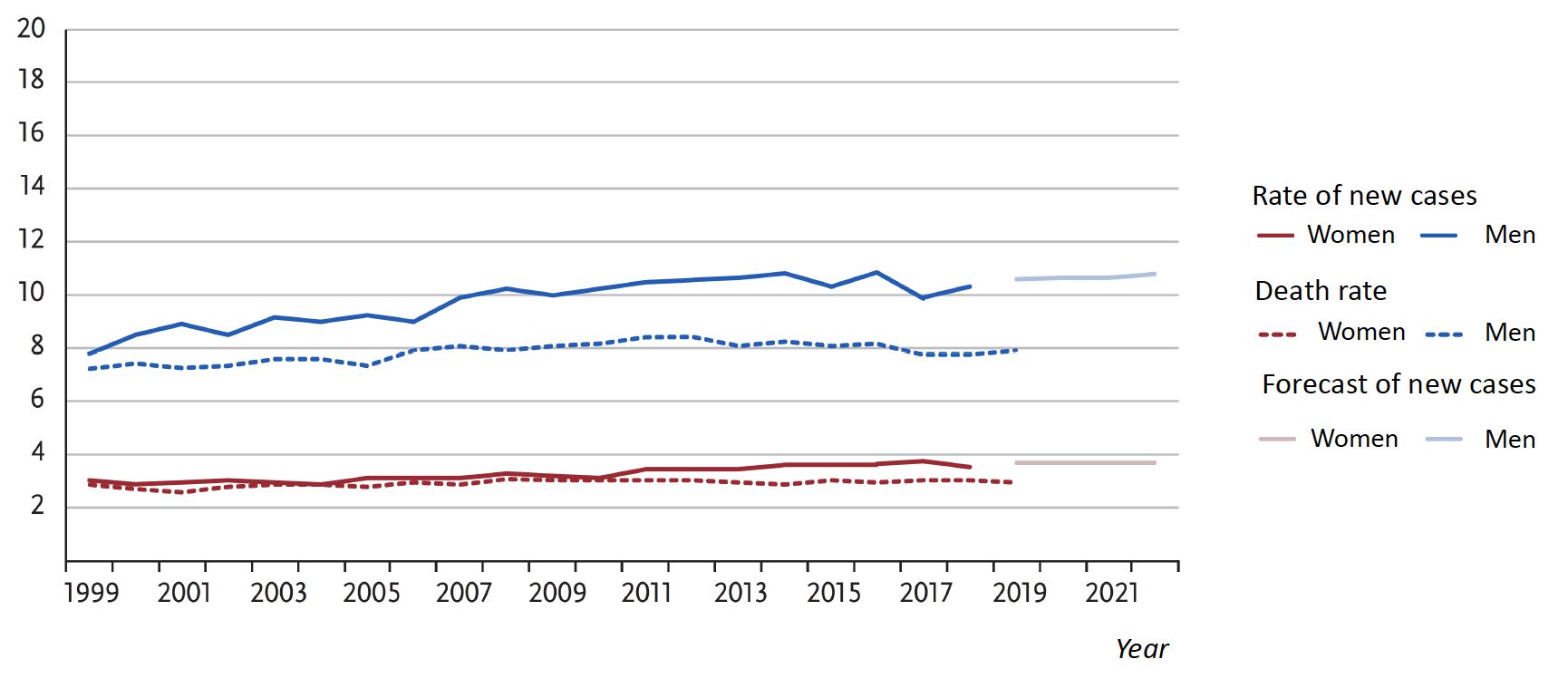
With approximately 6,000 new cases recorded annually in cancer registries, HCC is the most common malignant tumor of the liver in Germany. About three-quarters of cases occur in men. According to cause-of-death statistics, approximately 4,300 deaths per year have been attributed to HCC in recent years. Age-standardized incidence and mortality rates have recently shown a slight decline among men, whereas they have remained unchanged among women (Figure 1).
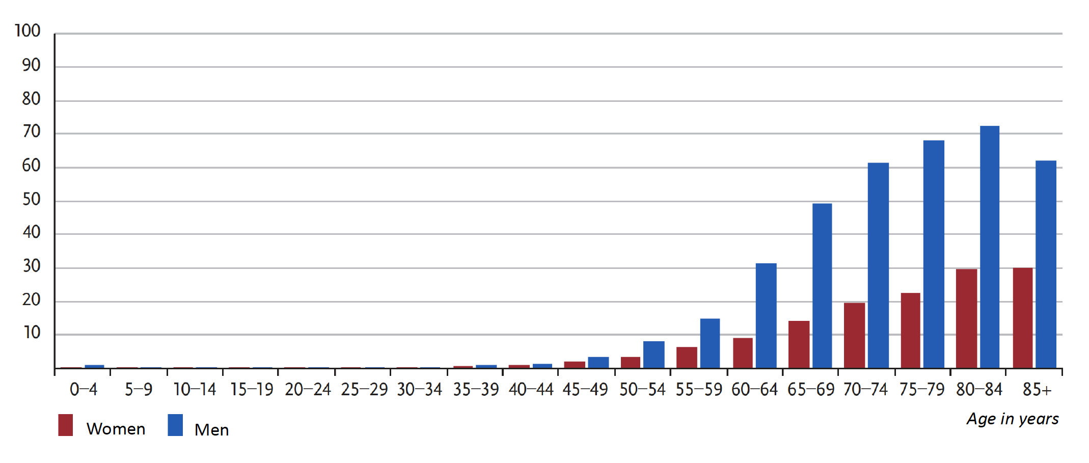
The median age at diagnosis is 71 for men and 74 for women. Figure 2 shows the current incidence rates in Germany by age and sex.
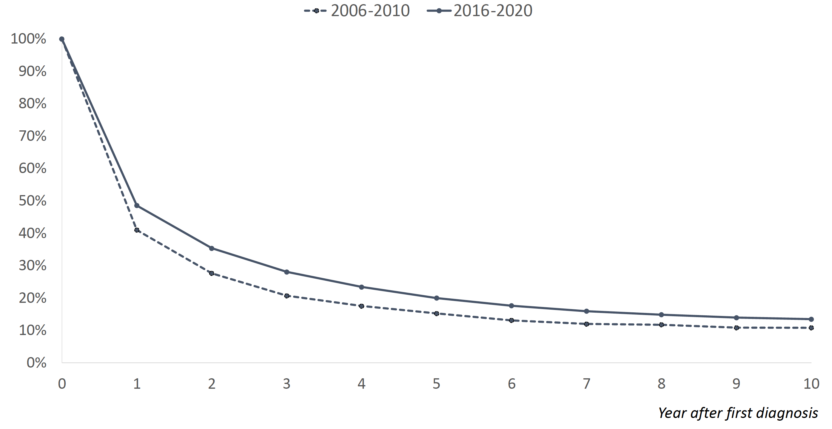
The median overall survival (OS) was most recently (2016–2020) 13 months for those under 60, 12 months for those aged 60 to 74, and 8 months for those over 75 years of age. The relative survival rates—which compare the observed OS to survival in the general population of the same age and sex—are 20% at 5 years and 13% at 10 years. The relative 5-year OS has thus increased by about 5% over the past 10 years.
According to epidemiological data from GLOBOCAN, 905,700 new cases of primary liver cancer and 830,200 deaths were recorded worldwide in 2020 [70], with cholangiocarcinomas included in these figures. The incidence has been rising globally in recent years. Up to 80% of global cases emerge in Southeast Asian countries and sub-Saharan African countries. The high incidence of chronic hepatitis B virus infection plays a decisive role here.
2.2Risk Factors
Liver cirrhosis is considered the most important risk factor for the development of HCC; in Germany, this is primarily due to excessive alcohol consumption and/or chronic hepatitis C. The annual risk of developing HCC in patients with existing liver cirrhosis is 2.5% per year [12]. However, this rate varies depending on the underlying cause and is 2% for hepatitis B-related cirrhosis and 3–8% for hepatitis C-related cirrhosis. For non-alcoholic steatohepatitis (MASLD, previously referred to as NAFLD) and non-alcoholic steatohepatitis (MASH, previously NASH), rates ranging from 0.004% to 7.6% have been reported [94].
In cases of chronic hepatitis B or C, as well as in MASLD, the risk of developing HCC exists even in the absence of cirrhosis and is 0.12% and 1.3%, respectively.
The underlying risk factors for primary hepatocellular carcinoma vary widely worldwide [31]. For example, alcohol consumption is the cause in 32% of cases in Western Europe, 53% in Eastern Europe, but only 13% in North Africa and the Middle East. In Latin America and West Africa, chronic hepatitis B is the dominant cause, accounting for 45% in each region, while in Western Europe, North Africa, and the Middle East, chronic hepatitis C is the leading cause of HCC, accounting for 44% in each region, and in the Asia-Pacific region, it accounts for as much as 55%. MASH and MASLD are significantly on the rise in Europe, as well as in the United States and China, as causes of liver cirrhosis and HCC [61, 94].
Patients with hemochromatosis have a 1.8-fold increased risk of HCC compared to patients with other forms of chronic liver disease [28].
In addition to nutritional and infection-related causes, genetic polymorphisms in the germline play a role in the risk of developing HCC. For example, the phospholipase PNPLA3 variant rs738409 and the TM6SF2 variant rs58542926 are associated with an increased risk of HCC in patients with alcohol-related liver cirrhosis. In contrast, a polymorphism at locus rs2242652(A) of the telomerase reverse transcriptase TERT is associated with relative protection against the development of HCC [87, 12]. Various polygenic risk scores have been developed to estimate the risk of HCC developing in a MASH/MASLD context [8, 52]; these scores can describe the probability of HCC developing from cirrhosis depending on the etiology of liver damage and the specific population studied (Asian vs. non-Asian).
TP53, TERT, and the activation of the hepatic WNT signaling pathway are considered molecular pathogenic drivers of HCC development [74, 87].
3Prevention and Early Detection
3.1Prevention
Key measures for reducing HCC risk in Western European countries are listed in Table 1.
A. Prevention of Cirrhosis (Proven Preventive Measures) |
|
|
|
B. Prevention of HCC (not established—retrospective data) |
3.2Early Detection
For the early detection of HCC, regular screening is recommended for patients with advanced liver fibrosis, such as those with chronic HCV infection or MASLD, as well as for patients with rare predisposing hereditary disorders such as acute intermittent porphyria, hereditary hemochromatosis, glycogen storage disease, Gaucher disease, or tyrosinemia type I [3].
For patients with liver cirrhosis, an HCC screening program involving qualified ultrasound plus AFP measurement every 6 months is recommended. Regular measurement of AFP levels appears appropriate, as AFP levels ≥ 20 ng/ml indicate HCC < 5 cm with a sensitivity of 49–71% and a specificity of 49–86% [80]. For screening patients with alcohol-related liver disease, MASLD, or cirrhosis following cured HCV infection, a lower AFP cutoff of 10 ng/ml has recently been advocated [111]. At the same time, a retrospective Korean analysis of >185,000 HCC patients showed that regular AFP testing improved OS [56]. This effect was particularly pronounced in patients with hepatitis B. However, a prospective phase III study demonstrated that the use of the GALAD score (gender, age, AFP L3, AFP, and Des-γ-carboxyprothrombin) or the GAAD score (excluding AFP L3) instead of AFP measurement alone at 6-month screening intervals significantly improved early HCC detection in patients with cirrhosis [109, 115]. Other complex scores, such as HES-V2, have already been validated in phase III studies in comparison to the GALAD score, among others [101].
Depending on the quality of the available ultrasound, switching to MRI—which generally has a higher sensitivity—is also recommended [102].
A randomized trial demonstrated an improvement in the early detection of HCC, as well as in surgical operability and OS, through screening [95]; these results were confirmed in a meta-analysis of 59 studies involving 145,396 patients [78]. Through structured screening of patients with liver cirrhosis, nearly twice as many patients were detected at an early stage of HCC (hazard ratio [HR] 1.83) and treated with curative intent (HR 1.83). This also had a significant impact on OS (HR 0.67) [78].
In patients with hemochromatosis, HCC can develop even in the absence of cirrhosis; therefore, screening is recommended as soon as the extent of liver fibrosis reaches a certain severity level (METAVIR F3, Ishak Stage 4–5) [24].
Patients with chronic hepatitis B and a non-cirrhotic liver represent a special case. In this group, the PAGE-B score was validated as a prognostic predictor for Caucasian patients [57] (Table 2). A PAGE-B score of <10 had a negative predictive value of 99% for the occurrence of HCC within the next 5 years. For patients with HBsAg-positive hepatitis B and cirrhosis, the AASLD guidelines recommend ultrasound and AFP testing every 6 months.
Age in years (points) | Gender (points) | Platelet count (points) | |||
|---|---|---|---|---|---|
16–29 | 0 | Female | 0 | >200/nl | 0 |
30–39 | 2 | Male | 6 | 100–199/nl | 6 |
40–49 | 4 | <100/nl | 9 | ||
50–59 | 6 | ||||
60–69 | 8 | ||||
>70 | 10 | ||||
4Clinical characteristics
In the early stages of HCC, when curative treatment options are available, patients usually do not show any specific symptoms. In developed healthcare systems, the diagnosis is therefore typically made as part of the recommended surveillance for cirrhosis or other severe chronic liver disease. Clinical signs of advanced disease are summarized in Table 3.
5Diagnosis
To confirm a diagnosis of HCC, imaging with pathognomonic findings and histopathological examination are available (Figure 4). Histological confirmation is required in all cases prior to the initiation of palliative therapy, as well as in potentially curative situations where the contrast agent behavior remains unclear in two independent imaging studies [87]. If a primarily surgical treatment plan with curative intent is decided upon, histopathological confirmation can be performed on the resected tumor tissue. For patients who are eligible for a liver transplant, the LI-RADS criteria are preferred, and a biopsy should be performed only in cases of unclear findings (LR3 or LR4) to rule out a mixed tumor (HCC/cholangiocarcinoma) [73]. In patients with suspected HCC who do not have liver cirrhosis, histopathological confirmation is required [31].
5.1HCC Criteria in Diagnostic Imaging
Diagnostic categories for confirming the diagnosis through imaging using dynamic MRI (preferably with hepatocyte-selective contrast agent [117, 120, 129]) according to the Liver Imaging and Reporting System (LI-RADS) include tumor size, contrast agent kinetics (arterial and washout phases), capsular enhancement, and growth dynamics (≥50% increase in ≤6 months, ≥100% increase in >6 months, new mass ≥10 mm). These criteria result in the LI-RADS categories [38], which are currently applied in their revised version (rLI-RADS) [34].
Despite the high diagnostic accuracy, however, approximately 9% of cases result in false-negative assessments when imaging alone is used [18]; therefore, histological confirmation should be performed, particularly in patients who are to receive systemic therapy.
5.2Histopathological Evaluation
The histopathological classification of HCC should be based on the current WHO classification (most recently updated in 2019) [51], which requires a biopsy. The diagnosis is based on defined histomorphological criteria for hepatocellular differentiation (trabecular growth, bile production, medium- to large-sized cells with round nuclei and prominent nucleoli) and malignancy (architectural disruption with reduction of the reticulin fiber network, nuclear atypia, vascular invasion). Specific subtypes (steatohepatitic, clear-cell, macrotrabecular, cirrhotic, neutrophil-rich, lymphocyte-rich, chromophobic, fibrolamellar) can be identified based on their characteristic morphology, in some cases on molecular alterations (e.g., the DNAJB1-PRKACA fusion in fibrolamellar HCC), and on the appearance of the non-tumorous liver parenchyma. Immunohistochemical expression of arginase-1 and HepPar1 is helpful in distinguishing non-hepatocellular tumors, such as metastases, and in determining the lineage differentiation in combined HCC-cholangiocarcinomas. A particular challenge in biopsy is distinguishing highly differentiated HCC from benign, preinvasive, and early hepatocellular lesions. These include focal nodular hyperplasia (FNH), hepatocellular adenoma, dysplastic nodules, early-stage HCC (<2 cm in diameter, well-differentiated, non-encapsulated), and small, progressive HCC, in which intra- and extrahepatic metastases may occur [35]. In such cases, an immunohistochemical panel using the antibodies glypican-3, HSP70, and glutamine synthetase may be helpful [87]. If ≥2 of these markers are positive, the specificity for an HCC diagnosis is 100% [22, 82]. The detection of mutations in the hTERT promoter also supports the diagnosis of HCC.
The histopathological findings of a resected specimen or explant should include the extent of the tumor (staging) according to the current TNM classification, its type and degree of differentiation (grading), and the presence or absence of tumor tissue at the resection margin (R classification). The template from the International Collaboration on Cancer Reporting (ICCR) is recommended for standardized reporting [58]. Grading has prognostic relevance following resection and transplantation; a three-tiered system is currently recommended.
5.3Molecular Pathological Tests
Molecular testing is not (yet) necessary for the treatment of HCC. However, molecular pathological methods can be used to determine the entity and grade of the tumor. Molecular testing to identify potential therapeutic targets is not standard practice for curably treatable HCC, but may be useful as an individualized therapeutic attempt or for inclusion in clinical trials. Potential targets for molecularly targeted systemic therapies are summarized in Table 4 (according to [54]).
Molecular pathological methods can be used to support tumor typing and grading of hepatocellular tumors.
In analyses of cell-free DNA from circulating blood, molecular alterations were detected in 92.2% of cases, and potential therapeutic targets (TSC1/-2 18%, BRCA1/-2 8%, and PIK3CA 8%) were identified in 37% of cases [19].
A specific fusion transcript (DNAJB1-PRKACA) is pathognomonic for fibrolamellar HCC and is also being evaluated as a target for molecularly targeted therapy [4].
Typically, molecular sequencing analyses in HCC reveal a low to moderately increased tumor mutational burden (TMB) with an average of 2.9 mutations per megabase (Mut/Mb), corresponding to approximately 40–60 coding somatic mutations. Recurrent genetic alterations include TERT promoter mutations (50–60%), TP53 alterations (20–40%), CTNNB1 mutations (15–40%), and ARID1A mutations (10–20%) [87]. In HCC of non-viral etiology, EGFR/FGFR/RAS/MAPK alterations are most commonly found [54, 118].
Activation of oncogenic signaling pathways (Wnt-TGFβ, PI3K-AKT-mTOR, RAS-MAPK, MET overexpression, IGF) is frequently detected, as is FGF19/FGFR4 overexpression [42], which potentially allows for the targeted use of FGFR4 inhibitors.
Determination of MSI/MMR status, TMB, or PD-L1 expression has not yet been established as a routine parameter for the initial diagnosis of HCC. The use of immunotherapy regimens is not currently stratified based on these findings (see chapter 6: Treatment). Routine testing for NTRK fusions is also not indicated for primary diagnosis. However, testing for these molecular pathological alterations may be indicated in the context of deciding on systemic therapy after established standard options have been exhausted. In the presence of a (very rare) NTRK fusion or a TMB >10 Mut/Mb, reference can be made to existing tumor-agnostic approvals for entrectinib, larotrectinib, or pembrolizumab.
Testing for germline mutations is not currently recommended as a routine procedure [32, 53].
Genetic variant | Patients with HCC (n = 2,372) | Potential therapy |
|---|---|---|
FGF19 | 5.7% | Irpagratinib (ABSK-011) Lenvatinib |
TSC2 | 4.8% | mTOR inhibitors (e.g., everolimus) |
TMB-H | 3.4% | Immune checkpoint inhibition (ICI) |
BRCA1/BRCA2 | 2.2% | PARP inhibitors such as olaparib |
MET | 1.8% | Capmatinib Tepotinib |
CCNE1 | 1.6% | Lunresertib (RP-6306) |
ERBB2 (HER2) | 0.9% | Trastuzumab Trastuzumab deruxtecan Trastuzumab/Tucatinib Zanidatamab Trastuzumab/Pertuzumab |
MDM2 | 0.5% | Brigimadlin (development discontinued in 2025) |
FBXW7 | 0.5% | Lunresertib |
EGFR | 1.0% | Lenvatinib-based therapy |
BRAF | 0.4% | Encorafenib Vemurafenib Dabrafenib |
TP53Y220C | 0.8% | Rezatapopt (PC14586) |
MSI-H | 0.1% | ICI |
NTRK | 0.1% | Larotrectinib Entrectinib |
ROS1 | 0.1% | Crizotinib Entrectinib Repotrectinib |
KRAS
|
0.08% 0.1% |
Sotorasib Adagrasib Daraxonrasib Setidegrasib (ASP3082) Zoldonrasib (RMC-9805) |
FGFR2 | 0.3% | Pemigatinib Futibatinib Erdafitinib Lirafugratinib Tinengotinib |
ALK | 0.04% | Crizotinib Entrectinib Alectinib |
RET | 0.04% | Selpercatinib |
5.4Staging
Staging of HCC should include a contrast-enhanced CT scan of the thorax and abdomen. If the contrast-enhanced MRI provides diagnostic coverage of the entire abdomen, only a native CT scan of the thorax should be performed. With regard to the morphological aspects of the tumor, imaging analysis methods that take vascularity into account should be used [3]. To assess possible vascular infiltration, which is an important prognostic factor, a contrast-enhanced MRI using gadobutrol (Gadovist®) is recommended. In subsequent evaluations, a liver-specific contrast agent is preferred.
Staging serves to determine the TNM classification, the resulting stage assignment (currently according to the AJCC 8th edition, 2017), the tumor grade and degree of fibrosis, as well as to determine the BCLC stage according to the Barcelona criteria [68] (Table 5). To assess treatment options, it is necessary to determine hepatic functional reserve in cases of liver cirrhosis using the Child-Pugh score [62] (Table 6). Focusing solely on the BCLC stage does not reflect current clinical practice when making treatment decisions. Several studies have shown that the decision made by the multidisciplinary tumor board at the respective center regarding the appropriate therapy for the patient may yield better success rates than a decision based solely on the BCLC score [33, 47].
The indication for liver transplantation (LTx) exists provided that macrovascular invasion and extrahepatic tumor manifestations are ruled out and the patient is generally eligible for LTx. The assignment of a match-MELD score via standard exception [48, 88] is based on the Milan criteria [49], whereby tumors smaller than 20 mm are to be surgically resected, and thus the Milan criteria have been replaced by UNOS T2. The static classification serves solely to prioritize patients on the waiting list.
It is becoming increasingly evident that the dynamic selection criteria (biological response, AFP slope, G3, V+) are more suitable than the static ones.
Stage | Definition |
|---|---|
Very early stage (0) | Single liver lesion ≤ 2 cm Preserved liver function, general condition ECOG 0 |
Early stage (A) | Single or up to 3 liver lesions, each ≤ 3 cm Preserved liver function, ECOG performance status 0 |
Intermediate stage (B) | Multiple liver lesions Preserved liver function, ECOG performance status 0 |
Advanced stage (C) | Portal invasion and/or extrahepatic spread Preserved liver function, general condition ECOG 1–2 |
Terminal stage (D) | Any tumor spread End-stage liver failure, general condition ECOG 3–4 |
Clinical/biochemical parameters | Score points for increasing abnormality | ||
|---|---|---|---|
1 | 2 | 3 | |
Severity of encephalopathy | None | 1–2 | 3–4 |
Ascites | Absent | Mild | Moderate |
Albumin (g/dL) | Over 3.5 | 2.8–3.5 | Less than 2.8 |
PTT seconds above normal INR | Less than 4 Below 1.7 | 4–6 1.7–2.3 | More than 6 Over 2.3 |
Bilirubin (mg/dL) - in primary biliary cirrhosis | Less than 2 Less than 4 | 2–3 4–10 | Over 3 Over 10 |
Class A: Low surgical risk; Class B: Moderately increased surgical risk; Class C: High surgical risk
6Treatment
6.1Basic Principles
To determine the appropriate treatment approach, all patients with HCC should be presented at a multidisciplinary tumor board at a center affiliated with a liver transplant center. This requires the participation of qualified specialists from the disciplines of radiology (diagnostic and interventional), radiation oncology, nuclear medicine, pathology, gastroenterology/hepatology, visceral surgery, and hematology/oncology. To make an informed decision, the following information may need to be considered: histopathological findings, radiological findings, infection status (hepatitis), tumor burden (stage and TNM classification according to UICC), BCLC stage, current liver function parameters, AFP level, platelet count, Child-Pugh/ALBI stage in cases of liver cirrhosis, and general condition (Karnofsky or ECOG performance score) may need to be available.
For patients with chronic HBV infection and HCC, guideline-based antiviral therapy is indicated. Tenofovir and entecavir are established as the standard of care in this setting. The indication for antiviral therapy also applies to patients with chronic HCV infection and HCC, adjusted as necessary based on the HCV genotype and the corresponding approval status [59]. In curative-intent treatment, antiviral therapy for HCV infection has been shown to improve OS [13, 59]. This study demonstrates the effectiveness of HBV therapy in reducing late HCC relapses following curative therapy [99].
1 LTx – Liver transplantation
2 Combination of local and systemic therapy, if appropriate
6.2Liver Transplantation
It is recommended that a liver transplant center be consulted for the initial decision-making process in cases of HCC amenable to curative treatment. This also applies to patients with resectable HCC; however, it is particularly relevant for patients with unresectable HCC and cirrhosis who meet the Milan criteria (BCLC A), but may also apply to patients with surgically resectable or borderline resectable HCC and cirrhosis who meet the Milan criteria [49] [3] (see Table 7). The indication and urgency for a liver transplant must therefore be determined as quickly as possible at a liver transplant center. A transplant may also be indicated outside the UNOS T2 criteria (see Table 7). In Germany, listing based on “Standard Exceptions” (SE) criteria is not possible for these tumors; therefore, other listing options must be evaluated with the respective transplant center (living donation, listing without SE criteria, center-specific offers). This applies in particular to tumors that have responded very well to local and systemic therapy. The level of AFP correlates with transplant outcomes [6], and an AFP level >1,000 ng/ml constitutes a contraindication for liver transplantation [5]. A decrease to < 500 ng/ml (preferably <200 ng/ml) achieved through locoregional or systemic therapy leads to an improved prognosis after transplantation [50].
In patients who are initially ineligible for transplantation but who receive first-line systemic therapy with atezolizumab + bevacizumab, liver transplantation may still be indicated in approximately one-quarter of patients who respond well [133]; in such cases, the patient should be referred back to a liver transplantation center.
When comparing patients who received a liver transplant while meeting the Milan criteria with those who met the Milan criteria only after downstaging, the 10-year OS and the relapse rate are comparable (61.5% vs. 52.1% and 13.3% vs. 20.6%, respectively) [81].
According to the guidelines of the German Medical Association, a de novo HCC may be assumed if the recurrence-free interval exceeds 2 years, which may result in an indication for LTx.
Liver transplantation is not indicated in cases of extrahepatic HCC manifestations and/or macrovascular invasion of the hepatic vessels.
If liver transplantation is indicated, bridging therapy using locoregional treatment (TACE/ablation (see below) or surgical resection) should be pursued. The decision to initiate bridging therapy should always be made in consultation with a transplant center.
Outside of clinical trials, patients with HCC should not receive adjuvant systemic therapy following liver transplantation. This does not preclude the continuation of antiviral therapy for HBV- or HCV-related HCC (see above), which should be decided upon through a multidisciplinary approach.
|
|
|
|
|
|
|
|
|
6.3Primary Surgical Approach With or Without Neoadjuvant/Adjuvant Therapy
A prerequisite for primary surgical resection is the possibility of a complete (R0) surgical resection. Portal hypertension (splenomegaly, esophageal varices, ascites, thrombocytopenia) should be ruled out beforehand, if indicated by measuring wedge pressure.
If not all intrahepatic HCC lesions can be completely (R0) surgically resected, a decision should be made prior to treatment regarding the combination with local ablation or embolization procedures (see below) with curative intent.
A retrospective multicenter study from Italy [122] involving 720 patients with early-stage multinodal HCC showed, in a “matched-adjusted” comparison between primary liver resection (n=296), percutaneous RFA (n=240), and TACE (n=184), advantages in 3- and 5-year OS in favor of primary liver resection (3-year OS 71% vs. 65% vs. 49%, 5-year OS 56% vs. 40% vs. 29%).
Liver resection should be performed for a solitary HCC nodule <2 cm in patients with liver cirrhosis who are functionally resectable. For tumors >2 cm, an individualized discussion should take place, in which the tumor’s location, tumor biology, risk of recurrence, and a potential living donor option should be factored into the decision. Liver resection can be performed via open or minimally invasive surgery. If the resection is performed as a bridging procedure to a planned liver transplant, it should be conducted using a minimally invasive approach.
If neoadjuvant or adjuvant systemic therapy is being considered before or after an R0 resection, enrollment in clinical trials is recommended. The current data on this are as follows:
For adjuvant therapy, the first interim analysis of the treatment with atezolizumab plus bevacizumab in a randomized comparison with follow-up alone (phase III IMbrave 050 trial) demonstrated a significant improvement in progression-free survival (PFS, primary endpoint) [64]; however, this benefit could not be confirmed with longer follow-up. Although the study was not powered for OS, the mature OS data [93] showed no benefit of the treatment compared to follow-up alone; therefore, treatment with atezolizumab plus bevacizumab should not be used as adjuvant therapy.
Similarly, the adjuvant administration of pembrolizumab in patients with a complete radiological response in the phase III KEYNOTE-937 trial showed no benefit in the primary endpoint of PFS [98].
Perioperative systemic therapy with nivolumab or nivolumab plus ipilimumab has been shown to be safely feasible; histopathologically, a major pathological response was documented in individual patients following neoadjuvant administration [36]. As of June 2026, there is no EMA approval for this indication.
In a randomized trial comparing perioperative systemic therapy with camrelizumab plus rivoceranib (also known as apatinib) to surgical therapy alone in 294 patients with Child-Pugh A and primarily resectable HCC, median event-free survival (EFS) improved from 19 to 42 months [124]. There is no EMA approval for this indication; camrelizumab is not yet approved in Germany (as of June 2026).
6.4Local Ablative Procedures
6.4.1Potentially Curative Approach
Patients undergoing a primary local ablative treatment regimen have an overall curative potential of 20–30%, and up to 40% for small, solitary HCC lesions (n=1,571) [20], whereby percutaneous ablation of the HCC should be performed using radiofrequency ablation (RFA) or microwave ablation (MWA).
In patients with HCC up to 3 cm in size, surgical resection and ablation are equivalent procedures in terms of clinical outcomes [110]. The indication for primary thermal ablation applies in particular to HCC ≤ 3 cm located in a site unfavorable for resection or in cases of significantly impaired liver function.
The advantages of percutaneous MWA include low associated morbidity—particularly with regard to post-procedure pain—a short hospital stay, and the option of performing the procedure under sedation rather than general anesthesia.
In patients with an HCC lesion >3 cm and ≤5 cm who have good liver function (Child-Pugh A) and mild or moderate portal hypertension, TACE should be performed prior to thermal ablation [3].
In recent years, several guidelines (including those from the German AWMF, NCCN, ESMO, and EASL) have included “stereotactic body radiotherapy” (SBRT) as a local ablative procedure. In SBRT, very high, ablative radiation doses are delivered over a few sessions (usually 3–10). The advantages of SBRT include its feasibility in an outpatient setting and the non-invasive nature of the therapy. A meta-analysis of 1,889 patients demonstrated a three-year local control rate of 84% for this procedure [16]. In the case of a solitary HCC relapse ≤ 5 cm, a single-center prospective randomized trial involving 166 patients showed that SBRT was more effective than RFA in terms of local PFS (HR 0.45, p=0.014). The 2-year survival rates and the adverse side effects of the treatment did not differ significantly [127]. Several studies have now also demonstrated promising data regarding local control for the use of SBRT as a bridging procedure in the context of a planned transplant. For example, local control one year after SBRT was 100% in the study by Lee et al. and 92.3% in the study by Wong et al. [45, 126].
Downstaging with the goal of resection or liver transplantation is also increasingly being considered as a treatment strategy and applies to patients in advanced stages, including those with a high tumor burden or portal vein invasion [125]. The primary consideration in selecting the appropriate treatment modality is an objective response while preserving liver function. In a multicenter cohort of 326 patients meeting UNOS downstaging criteria compared to 190 “all-comers” (any tumor burden, no extrahepatic metastases), 82% and 66%, respectively, achieved successful downstaging to meet UNOS transplantation criteria. Y90 radioembolization (SIRT/TARE) was superior to chemoembolization in this regard (p<0.001) [128]. In clinical practice, the appropriate sequence and selection of locoregional procedures and systemic therapy (particularly immunotherapy) are likely to be of crucial importance and should be determined through a multidisciplinary approach. Currently, the situation is complicated by the fact that hardly any data beyond a “proof-of-concept” are available.
6.5Local Treatment Approaches in the Intermediate Stage
In the intermediate stage, for unresectable HCC, the indication for intra-arterial therapeutic procedures should first be evaluated. TACE and TARE (or SIRT) are available as locoregional therapeutic options. Both TACE and SIRT/TARE must be administered selectively and in a tumor-targeted manner to preserve the patient’s liver function [107], TACE is typically performed repeatedly, although repeat procedures should be contingent upon evidence of an objective response and the preservation of liver function [11, 86]. This decision should be made by a multidisciplinary tumor board and reevaluated no later than after two treatments. Meta-analyses have found the two procedures to be equivalent in terms of OS; however, TARE was significantly superior to TACE in terms of time to progression [10]. A monocenter, prospective study comparing TARE with TACE in the intermediate stage serves as an example [100]. The superior TTP of TARE compared to TACE (HR 0.36) led to the study’s termination following a planned interim analysis after 72 patients. TACE maintained its superiority in both tolerability and OS. Such consistent data from different studies are particularly important, especially in the bridging phase to transplantation.
In the results of the EMERALD-1 study, first presented in 2024, a significant advantage of the combination of TACE with durvalumab and bevacizumab compared to TACE alone was observed in 616 HCC patients with Child-Pugh A to B7 in terms of PFS (HR 0.77, 95% CI 0.61–0.98, p=0.032), but no benefit in OS [40], even in the longer-term data analysis [132], so this treatment modality does not represent a new standard of care. Similarly, the randomized comparison of lenvatinib + pembrolizumab + TACE versus placebo + TACE (LEAP-012) in 480 HCC patients with Child-Pugh A in first-line therapy showed a significant PFS benefit for the combination therapy (HR 0.66, 95% CI 0.51–0.84; p=0.0002) [44], but no OS benefit. The EMERALD-3 study (STRIDE + TACE vs. STRIDE + TACE + lenvatinib vs. TACE alone) demonstrates a clear PFS benefit and a potential OS benefit—which has not yet been confirmed—for the combination arms [131]. These data should be discussed with patients in the intermediate stage who have adequate liver function and a good general condition, and may represent a strategy for bridging to a liver transplant (LTx).
6.6Systemic Therapy
Systemic drug therapy using tyrosine kinase inhibitors (TKIs; sorafenib, lenvatinib, regorafenib, or cabozantinib) or immunotherapies (atezolizumab, durvalumab, pembrolizumab, tislelizumab, nivolumab, ipilimumab, tremelimumab) or anti-angiogenic antibodies (bevacizumab, ramucirumab, sometimes in combination therapies) has been established as the standard of care since the publication of the results of the SHARP study [42]. While the SHARP trial compared sorafenib with placebo, subsequent studies compared the new therapeutic approach against sorafenib and/or lenvatinib as the control arm. For ethical reasons, no further randomized comparisons with placebo were conducted; however, no randomized comparisons of newer therapeutic approaches against one another were performed either, which prevents a nuanced assessment of the benefits of the numerous newer treatment options.
In principle, for patients for whom the liver transplant center does not consider a curative treatment option to be available, treatment with atezolizumab plus bevacizumab, durvalumab with or without tremelimumab, nivolumab with ipilimumab, as well as with sorafenib, lenvatinib, regorafenib, cabozantinib, and ramucirumab is possible. For patients who are initially ineligible for transplantation and who are treated with first-line systemic therapy consisting of atezolizumab plus bevacizumab, liver transplantation may still be indicated in approximately one-quarter of patients who show a good response [133]; in such cases, the patient should be referred back to a liver transplant center.
For Child-Pugh stage B, data are available from observational studies for sorafenib and from smaller phase II studies for ICI. A meta-analysis of previously published reports on PD-1 antibodies in patients at this stage confirmed an acceptable overall safety profile for these agents, although with higher associated morbidity than in patients with Child-Pugh A cirrhosis [89]. Similarly, in a retrospective analysis of 343 patients with Child-Pugh B (EU, USA, Asia) from 2017–2022, treatment with atezolizumab plus bevacizumab or nivolumab resulted in a significant improvement in median OS from 4 to 7.5 months compared to best supportive care (HR 0.59, p<0.001) [106]. Focusing on patients with Child-Pugh B7 (see Table 6), it was reported that the subgroup of patients with slightly increased bilirubin, moderately reduced albumin, mild ascites, and no hepatic encephalopathy can achieve a clinical outcome comparable to that of Child-Pugh A patients when treated with atezolizumab and bevacizumab [112]. Accordingly, systemic therapy may also be considered for selected patients with Child-Pugh B7 who are in good general condition (ECOG PS≤1) [3, 32, 107]. In patients with Child-Pugh C cirrhosis, systemic drug therapy for HCC is not indicated.
Systemic therapy should not be continued beyond the point at which treatment failure is confirmed; however, following a decision by a multidisciplinary tumor board, it should be switched to another systemic therapy if indicated.
If a patient with HCC that is not initially amenable to curative treatment, but without distant metastases, shows a very good response to systemic anticancer therapy, it is recommended that this case be presented again to the tumor board to discuss the possibility of secondary, potentially curative therapy [3].
6.7First-Line Systemic Therapy
The currently available study results on the above-mentioned agents for first-line systemic therapy in HCC can be summarized as follows:
In the combination of atezolizumab plus bevacizumab versus sorafenib, an overall response rate of 27.3% versus 11.9% was observed, along with a PFS of 6.8 versus 4.3 months (HR 0.66; p<0.001), and a 1-year OS of 67.2% vs. 54.6% [26]. Longer follow-up revealed a median OS of 19.2 vs. 13.4 months (HR 0.65; p<0.001) [69]. These results were also replicated under practical “real-world” conditions [29].
In a phase Ib/II study involving 59 patients with Child-Pugh A an improvement in response rate, PFS, and OS compared to atezolizumab-bevacizumab [104]; however, the prospective randomized IMbrave152/SKYSCRAPER-14 study showed no benefit compared to atezolizumab/bevacizumab alone [105].
Adding ipilimumab to the atezolizumab/bevacizumab combination did not result in an improvement in response in a randomized comparison with atezolizumab/bevacizumab (“TRIPLET HCC”) [116].
The combination of tremelimumab and durvalumab (“STRIDE” regimen) (HIMALAYA study) versus sorafenib yielded a response rate of 20.1% versus 5.1% (for the STRIDE regimen), a PFS of 3.8 versus 4.1 months (for sorafenib) (HR 0.90 for STRIDE) and an OS of 16.4 vs. 13.8 months (HR 0.76; p=0.0008) [1]. The analysis of patient-reported outcomes (PROs) showed an improvement in clinical symptom burden and quality of life for patients receiving tremelimumab plus durvalumab compared with sorafenib [119].
The combination of nivolumab plus ipilimumab (CheckMate-9DW study), tested against lenvatinib or sorafenib, showed a response rate of 36% vs. 13% in 668 patients, a PFS of 7.5 vs. 7.5 months (HR 0.72) and an OS of 23.7 vs. 20.6 months (HR 0.79; p=0.018) [21].
In the CARES-310 trial, camrelizumab plus rivoceranib (also known as apatinib) versus sorafenib achieved an improved PFS of 5.6 vs. 3.7 months (HR 0.52; p<0.0001) and significantly longer OS of 22.1 vs. 15.2 months (HR 0.62; p<0.0001). Overall, treatment-related toxicity was increased with this combination, although no negative impact on patients’ quality of life was observed [63].
Compared with sorafenib, lenvatinib demonstrated a PFS of 7.4 vs. 3.7 months and an OS of 13.6 vs. 12.3 months (HR 0.92) [39]. Similar to sorafenib, more recent phase III studies with lenvatinib achieved an OS of up to 20 months.
Compared with placebo, sorafenib demonstrated a response rate of 2.3%, progression-free survival (PFS) of 4.9 vs. 4.1 months, and a significantly improved OS of 10.7 vs. 7.9 months (HR 0.69; p<0.001) [42]. In recent phase III trials in which sorafenib served as the control arm, an OS of 13–15 months was achieved, which is likely attributable primarily to the use of evidence-based second-line therapies.
The combination of lenvatinib with pembrolizumab versus lenvatinib plus placebo in first-line therapy among 794 patients with Child-Pugh A (LEAP-002) did not result in a significant improvement in OS or PFS with the combination therapy [114].
In a comparison of the immunotherapy/TKI combination of atezolizumab plus cabozantinib with sorafenib, the COSMIC study showed a significantly prolonged PFS of 6.9 vs. 4.3 months (HR 0.63; p=0.0012), but comparable OS of 16.5 vs. 15.5 months (HR 0.90; p=0.44) [91].
Another option for first-line systemic therapy, toripalimab plus bevacizumab, was randomized against sorafenib in the phase III HEPATORCH trial. In 326 Asian patients with Child-Pugh A, the combination of the PD-1 antibody with bevacizumab resulted in a significant improvement in PFS and OS [121]. Toripalimab is approved in the EU for the treatment of nasopharyngeal and esophageal carcinoma.
Despite differences in study results—which are likely due primarily to study-specific selection criteria—meta-analyses show no significant difference between the various combination therapies tested [15, 30, 113].
Based on current data, first-line therapy with an ICI as monotherapy is not superior to sorafenib:
Durvalumab vs. sorafenib [1]—however, due to its non-inferiority to sorafenib, durvalumab is approved by the EMA as monotherapy for first-line treatment of HCC.
Tislelizumab vs. sorafenib [66]
Nivolumab vs. sorafenib [91]
The FDA’s previously granted accelerated approval of nivolumab for this indication was revoked in July 2021.
In a randomized phase III trial from China, the combination of lenvatinib with TACE demonstrated a significant improvement in OS (17.8 vs. 11.5 months) and PFS (10.6 vs. 6.4 months), as well as a better response rate according to mRECIST (54.1% vs. 25.0%) in patients with advanced-stage disease [60]. However, the use of local therapies for BCLC C is not considered an accepted standard of care [3] and should only be performed following a multidisciplinary discussion in the tumor board.
The combination of camrelizumab and rivoceranib (also known as apatinib) with TACE resulted in a significant improvement in PFS (3.2 vs. 10.8 months) compared to camrelizumab and rivoceranib alone in a randomized phase II trial (n=200 patients). OS was positively influenced by the combination therapy; however, the follow-up period is still too short and fewer than 50% of OS events have occurred, so a definitive assessment cannot yet be made [130]
In intermediate-risk patients with relapse following prior primary resection, a randomized phase III trial comparing local ablation with TACE alone versus a combination of sorafenib and TACE demonstrated a significant prolongation of median OS from 15 to 22 months (HR 0.55, p<0.001) was achieved with the combination therapy [103].
According to the results of the CATCH-IT study, treatment with ICI can be used in patients with HIV infection with comparable efficacy and safety to that observed in HIV-negative patients [23].
Consequently, for first-line systemic therapy in patients who cannot be treated curatively with Child-Pugh stage A cirrhosis (5–6 points; see Table 6) and no contraindications, the combination of atezolizumab plus bevacizumab, or durvalumab plus tremelimumab, or nivolumab plus ipilimumab should be used until radiologically detectable tumor progression occurs. Extending this recommendation to individual patients in Child-Pugh stage B7 requires a decision by the multidisciplinary tumor board on a case-by-case basis.
For patients with contraindications or intolerance to these agents, lenvatinib (or sorafenib) should be used as first-line monotherapy (see figure 5).
*Orphan drug status in the EU for this combination as first-line therapy for HCC; not yet approved in Germany
6.8Systemic Second-Line and Subsequent Therapies
Following failure of first-line systemic therapy, second-line therapy should be initiated. Phase III studies have demonstrated the efficacy of regorafenib, cabozantinib, and ramucirumab following failure of sorafenib therapy. For patients who have not been treated with sorafenib, no formal phase III data are currently available; however, treatment with the approved agents is recommended, particularly for patients with well-preserved liver function. To date, no randomized comparisons between the TKIs have been conducted for this indication. The addition of the PD-L1 checkpoint inhibitor atezolizumab to second-line therapy with lenvatinib or sorafenib did not demonstrate an OS benefit (primary endpoint) in the IMbrave251 study [123].
None of the aforementioned immunotherapies (atezolizumab, pembrolizumab, durvalumab, nivolumab, ipilimumab, tremelimumab) are approved for second-line therapy following TKI failure. If a patient did not receive immunotherapy-based treatment in the first-line setting (e.g., due to relative contraindications), this option may be considered in the treatment sequence. Studies conducted to date have yielded the following results:
In the Checkmate-040 study, the combination of nivolumab plus ipilimumab following prior treatment with sorafenib demonstrated a response rate of approximately 30% [90].
Pembrolizumab monotherapy, in a randomized comparison with placebo, resulted in a PFS of 3.0 vs. 2.8 months and an OS of 13.9 vs. 10.6 months [27].
In Asian patients who had previously received sorafenib or oxaliplatin-based chemotherapy, pembrolizumab monotherapy resulted in a response rate of 12.7% vs. 1.3%, a PFS of 2.6 vs. 2.3 months, and an OS of 14.6 vs. 13.0 months compared with placebo [65].
However, the VEGFR2 inhibitor ramucirumab is approved as monotherapy following sorafenib pretreatment for patients with an AFP level of ≥400 ng/mL. While no significant benefit was observed in this indication among unselected patients compared with placebo [97], a follow-up study demonstrated a significant OS benefit (8.5 vs. 7.3 months) in patients with a baseline AFP level of ≥400 ng/mL. PFS was also significantly prolonged, at 2.8 vs. 1.6 months [96].
The available TKIs (following failure of sorafenib) include lenvatinib and cabozantinib, regorafenib and ramucirumab (for AFP ≥400 ng/mL), and (following failure of lenvatinib) sorafenib and cabozantinib (see Figure 6). Lenvatinib is not approved for second-line therapy following sorafenib failure.
Second-line therapies are also generally restricted to patients with adequate general health (ECOG ≤1) and Child-Pugh A cirrhosis; such therapy may be considered for Child-Pugh B7 (7 points according to Table 6) only in selected individual cases.
Experimental treatment options that can be derived from molecular pathology findings—such as dostarlimab for MSI-H/dMMR, selpercatinib upon detection of a RET fusion, larotrectinib or entrectinib upon (extremely rare) detection of an NTRK fusion, or pembrolizumab or nivolumab plus ipilimumab in cases of increased tumor mutation burden [53]—provided that ICI has not been used previously—are case-by-case decisions outside established treatment standards that may be recommended by a molecular tumor board.
7Systemic Tumor Treatment Agents (alphabetical)
7.1Atezolizumab
Atezolizumab is a humanized IgG1 antibody targeting PD-L1 and belongs to the class of ICIs. It is approved for the treatment of HCC [27] in combination with bevacizumab, as well as for a broad spectrum of other malignant neoplasms. For use in HCC, evidence of PD-L1 expression is not required. As with other ICI targeting PD-1 or PD-L1, immune-mediated adverse effects such as hepatitis, pneumonitis, colitis, endocrinopathies, or skin reactions have been documented in clinical trials, as well as fatigue, which can be severe in some cases. There is a risk of exacerbation of a pre-existing autoimmune disease. No clinically significant pharmacological interactions with other active substances have been reported; however, the efficacy of atezolizumab is expected to be impaired following prior administration of immunosuppressive medications.
7.2Apatinib (see Rivoceranib)
See Chapter 7.13 Rivoceranib.
7.3Bevacizumab
Bevacizumab is a monoclonal, antiangiogenic antibody directed against the vascular endothelial growth factor (VEGF). The prescribing information for atezolizumab indicates approval in combination with bevacizumab for first-line treatment of HCC, whereas this indication is absent from the prescribing information for the various bevacizumab formulations. Adverse reactions (grade 3 or 4) that occurred in more than 5% of patients in the registration studies were hypertension and proteinuria. Rarer critical complications include arterial thromboembolic events and gastrointestinal perforations. The note in the guidelines regarding the risk of bleeding in the presence of esophageal varices must be observed.
Bevacizumab is proteolytically degraded in the body. It is not eliminated via the kidneys or the liver. Therefore, it is unlikely that other drugs will have a significant pharmacokinetic effect on the action of bevacizumab. Cases of microangiopathic hemolytic anemia have been reported in patients receiving combination therapy with bevacizumab and sunitinib; this is also noted in the prescribing information for bevacizumab.
7.4Cabozantinib
Cabozantinib is a multikinase inhibitor. In addition to the VEGFR1, VEGFR2, and VEGFR3 kinases, it also inhibits AXL and MET. Cabozantinib is approved for second-line treatment following failure of sorafenib in hepatocellular carcinoma (prolonged OS compared to placebo) as well as in renal cell carcinoma and differentiated thyroid carcinoma. In first-line therapy, no improvement in OS was observed when used in combination with atezolizumab compared to sorafenib [92]. The most commonly reported adverse effects in larger clinical trials of cabozantinib monotherapy were palmar-plantar erythrodysesthesia (17%), hypertension (16%), diarrhea (10%), and fatigue (10%).
Cabozantinib exhibits very high plasma protein binding. As a result, it can displace other drugs that are strongly bound to plasma proteins from their plasma protein binding sites. In the case of drugs with a narrow therapeutic index, this can lead to an increase in both desired and undesired effects if their metabolic and excretion pathways are simultaneously impaired. When cabozantinib is taken with a very high-fat meal, its oral bioavailability is increased by 57% compared to when taken on an empty stomach. Cabozantinib is metabolized primarily by CYP3A4. Concomitant treatment with cabozantinib and strong CYP3A4 inducers may reduce the systemic availability of cabozantinib and thus its clinical efficacy. Concomitant treatment with cabozantinib and strong CYP3A4 inhibitors may increase the incidence of adverse effects. Concomitant use of cabozantinib with drugs that are strong CYP3A4 inducers or CYP3A4 inhibitors should be avoided. Throughout the entire duration of treatment with cabozantinib, patients should avoid consuming grapefruit, grapefruit-like fruits (e.g., pomelo, bitter orange), and preparations containing them. Myelosuppression caused by cabozantinib, which occurs very frequently, may be exacerbated by the concomitant use of other myelosuppressive drugs. Since electrolyte disturbances have been observed very frequently during therapy with cabozantinib, concomitant treatment with cabozantinib and drugs that prolong the QTc interval may increase the risk of polymorphic ventricular arrhythmias, known as “torsade de pointes.” During treatment with cabozantinib, attention should be paid to a possible decrease in the levels of individual or all blood cell lines. Appropriate measures should be taken as necessary. Concomitant treatment with cabozantinib and drugs that prolong the QTc interval should be avoided. If this is not possible, care should be taken to maintain electrolyte balance, and the QTc interval should be monitored regularly. Taking cabozantinib may lead to bleeding, which can sometimes be severe. This risk is increased by the concomitant administration of cabozantinib with anticoagulants. During concomitant treatment with cabozantinib and anticoagulants, coagulation-related laboratory parameters should be monitored regularly. Cases of gastrointestinal perforation have been reported in clinical trials. This risk may be increased by the concomitant use of cabozantinib with substances known to carry a risk of gastrointestinal perforation. Concomitant treatment with cabozantinib and drugs associated with a risk of gastrointestinal perforation should be avoided.
7.5Camrelizumab
Camrelizumab is a humanized IgG4 monoclonal antibody targeting PD-1 that belongs to the class of ICIs. It has not yet been approved in the EU. Camrelizumab was evaluated in combination with rivoceranib (also known as apatinib) for first-line treatment of HCC compared to sorafenib in the CARES-310 study [63], where it demonstrated a benefit in OS. In early August 2024, the EMA granted “Orphan Drug” designation to this combination for first-line treatment of HCC. In monotherapy for HCC, the main treatment-related adverse effects reported were vascular skin reactions (up to 70%), proteinuria (23%), elevated transaminases and bilirubin (15–20%), and thrombocytopenia (15%).
Serious pharmacological interactions are not expected due to the properties of camrelizumab; no reliable data are currently available on this topic.
7.6Durvalumab
Durvalumab is a humanized IgG1 monoclonal antibody against PD-L1 and belongs to the class of ICIs. It is approved for the treatment of small-cell and non-small-cell lung cancer, as well as hepatocellular and biliary carcinomas. For HCC, it is approved as monotherapy and in combination with tremelimumab for first-line therapy. When used as monotherapy for HCC, severe treatment-related adverse events were reported in 8.2% of patients [1]. As with other ICIs, immune-mediated adverse events such as pneumonitis, colitis, endocrinopathies, skin reactions, hepatitis, pancreatitis, and others have been documented with the use of durvalumab. There is a risk of exacerbation of pre-existing autoimmune diseases. In addition, fatigue and gastrointestinal side effects are frequently reported. No clinically relevant pharmacological interactions with other active substances have been identified; however, the efficacy of durvalumab is expected to be impaired following prior administration of immunosuppressive medications.
7.7Entrectinib
Entrectinib is a potent inhibitor of neurotrophic tropomyosin receptor kinases (NTRK) A, B, and C and is approved for the treatment of NTRK-fusion-positive tumors as well as ROS1-mutated non-small cell lung cancer. Across three cross-entity studies, the following adverse effects were reported: taste disturbances, constipation, diarrhea, fatigue, confusion, elevated serum creatinine, dysesthesia, nausea, vomiting, arthralgia, myalgia, and weight gain, as well as isolated cases of severe neurotoxicity. QT prolongation may also occur.
Concomitant use of strong or moderate CYP3A inhibitors (e.g., ritonavir, saquinavir, ketoconazole, itraconazole, voriconazole, posaconazole, grapefruit, or bitter orange) should be avoided; if unavoidable, the dose of entrectinib should be reduced in accordance with the prescribing information. Concomitant use of strong CYP3A4/PGP inducers (e.g., carbamazepine, phenobarbital, phenytoin, rifabutin, rifampicin, St. John’s wort, apalutamide, ritonavir) should be avoided. Entrectinib may inhibit P-glycoprotein; however, no clinically relevant effects, e.g., on digitoxin, have been observed.
7.8Ipilimumab
Ipilimumab is a medication belonging to the class of ICIs. It blocks the inhibitory T-cell regulator CTLA-4, thereby enhancing the body’s own immune response. It is approved for first-line treatment of HCC, as well as for the treatment of melanoma, renal cell carcinoma, non-small cell lung cancer, malignant pleural mesothelioma, squamous cell carcinoma of the esophagus, and colorectal cancer. Except for melanoma, this approval is contingent upon combination with nivolumab. In the phase I/II CheckMate 040 study on HCC treatment in combination with nivolumab [90], treatment-related adverse events were reported in 70–94% of patients across three different dosage regimens. The most common events included skin reactions, gastrointestinal symptoms such as diarrhea, and immune-mediated inflammatory reactions or organ dysfunction. Interstitial pneumonitis occurred in 10% of patients.
No clinically relevant pharmacological interactions with other active substances were observed; however, administration of immunosuppressive medications prior to the start of ipilimumab therapy is expected to impair the efficacy of ipilimumab.
7.9Larotrectinib
Larotrectinib is a selective NTRK inhibitor approved for the treatment of NTRK-fusion-positive tumors. Clinical trials have reported the following side effects: fatigue, elevated liver enzymes, confusion/dizziness, constipation, nausea/vomiting, and constipation; in addition, less common side effects include muscle and joint pain, edema, headache, weight gain, hyperglycemia, and peripheral neuropathy.
Pharmacological interactions have been reported with inhibitors (e.g., atazanavir, clarithromycin, indinavir, itraconazole, ketoconazole, nefazodone, nelfinavir, ritonavir, saquinavir, telithromycin, troleandomycin, voriconazole, or grapefruit) as well as CYP3A and P-glycoprotein inducers (e.g., carbamazepine, phenobarbital, phenytoin, rifabutin, rifampin, or St. John’s wort). The prescribing information notes that if concomitant administration with a potent CYP3A4 inhibitor is necessary, the larotrectinib dose should be reduced by 50%.
7.10Lenvatinib
Lenvatinib is a multi-tyrosine kinase inhibitor that inhibits VEGFR1–3, FGFR1–4, PDGFR alpha, PDGF, KIT, and RET. It is approved for the treatment of renal cell carcinoma. From the phase III study comparing it with sorafenib in first-line treatment of HCC [39], the following adverse events were primarily reported: cutaneous erythrodysesthesia on the palms and soles, hypertension, gastrointestinal symptoms (e.g., diarrhea, constipation, nausea/vomiting, loss of appetite, and weight loss), hypothyroidism, and elevated liver enzymes. According to the prescribing information, proteinuria, aneurysms, aortic dissections, renal failure, CNS toxicity, fistulas/perforations, bleeding, arterial thromboembolism, impaired wound healing, osteonecrosis of the jaw, and cardiac dysfunction such as QT interval prolongation may also occur.
Myelosuppression caused by lenvatinib may be exacerbated by the concomitant use of other myelosuppressive drugs. Since prolongations of ventricular repolarization have been observed during treatment with lenvatinib, the concomitant administration of lenvatinib with drugs that prolong the QTc interval may increase the risk of polymorphic ventricular arrhythmias, known as “torsade de pointes.” Concomitant treatment with lenvatinib and drugs that prolong the QTc interval should be avoided. If this is not possible, electrolyte balance should be maintained, and the QTc interval should be monitored regularly. Bleeding also occurs very frequently with the use of lenvatinib. Concomitant treatment with lenvatinib and anticoagulants may further increase the risk of bleeding. When lenvatinib is used concomitantly with anticoagulants, coagulation-related laboratory parameters should be monitored regularly. Renal dysfunction, particularly acute renal failure, frequently occurs during treatment with lenvatinib. Concomitant administration of lenvatinib and drugs that interfere with the renin-angiotensin-aldosterone system (RAAS) may be associated with an increased risk of acute renal failure. When lenvatinib is administered concomitantly with drugs that affect the RAAS, renal function should be monitored regularly. The administration of corticosteroids or NSAIDs during therapy with lenvatinib should be avoided.
Gastrointestinal perforations have been observed during treatment with lenvatinib. The risk of this may increase with the concomitant use of lenvatinib and drugs that interfere with prostaglandin metabolism (e.g., NSAIDs, corticosteroids).
Lenvatinib is primarily metabolized via oxidation by aldehyde oxidase. N-demethylation via CYP3A4 and glutathione conjugation represent minor metabolic pathways. Therefore, neither CYP3A4 inhibitors nor CYP3A4 inducers have a significant effect on the systemic availability of lenvatinib.
7.11Nivolumab
Nivolumab is a monoclonal anti-PD-1 antibody and belongs to the class of ICIs. It is approved in combination with ipilimumab for first-line treatment of HCC, as well as for use as monotherapy or combination therapy for the treatment of a broad spectrum of malignant neoplasms. In a phase III study comparing first-line treatment of HCC with nivolumab versus sorafenib [91], the most commonly reported treatment-related adverse effects of nivolumab were fatigue, skin reactions, gastrointestinal symptoms (diarrhea, nausea, loss of appetite), and elevated transaminases. Other possible adverse effects include, among others, anemia, hypoalbuminemia, hyperkalemia, elevated liver enzymes, heart failure, elevated serum amylase, hyponatremia, elevated creatine phosphokinase, and renal dysfunction, as well as sometimes severe fever and interstitial pneumonia (immune-mediated pneumonitis), and immune-mediated liver or kidney inflammation and endocrinopathies.
As with other humanized monoclonal antibodies, there is no pharmacological interaction with the cytochrome P450 isoenzyme system or other drug-metabolizing enzymes. If immunosuppressive medications have been administered previously, the efficacy of nivolumab is expected to be impaired.
7.12Ramucirumab
Ramucirumab is a human IgG1 antibody that specifically binds to the vascular endothelial growth factor (VEGF) receptor-2. It is approved for second-line therapy following failure of sorafenib in patients with HCC and a serum AFP level of ≥400 ng/ml, as well as for adenocarcinomas of the stomach or the gastroesophageal junction, colorectal carcinomas, and non-small-cell lung carcinomas. In the placebo-controlled phase III REACH-2 trial evaluating second-line therapy for HCC [96], the most commonly reported treatment-related adverse events in the ramucirumab arm were fatigue, nausea/vomiting, loss of appetite, proteinuria, hypertension, bleeding tendency, peripheral edema, and diarrhea or constipation. To prevent infusion-related adverse reactions, premedication with an H1 antagonist is recommended.
Clinically relevant pharmacological interactions with other active substances are not described in the prescribing information and, as with other humanized monoclonal antibodies, are not expected.
7.13Rivoceranib (also known as apatinib)
Rivoceranib is a VEGFR2-targeted tyrosine kinase inhibitor that has not yet been approved in the U.S. or the EU. It was evaluated in combination with camrelizumab for first-line treatment of HCC compared with sorafenib in the CARES-310 study [63], where it demonstrated an advantage in OS. In early August 2024, the EMA granted “Orphan Drug” designation to this combination for first-line treatment of HCC (https://elevartherapeutics.com/2024/08/01/elevar-therapeutics-granted-orphan-designation/). Side effects reported from monotherapy studies primarily include hypertension, gastrointestinal symptoms such as nausea and vomiting, fatigue, hand-foot syndrome, and skin reactions.
The primary metabolic pathway is CYP3A4, so relevant pharmacological interactions with CYP3A4 inhibitors such as itraconazole or voriconazole and CYP3A4 inducers such as rifampicin or St. John’s wort are to be expected. A prescribing information document is not yet available in the EU.
7.14Selpercatinib
Selpercatinib is a highly selective RET kinase inhibitor. It is approved across tumor types for the treatment of RET-fusion-positive tumors as well as for RET-mutated thyroid carcinomas. The main adverse effects in the tumor-agnostic phase I/phase II study were hypertension and elevated liver enzymes, along with fatigue, proteinuria, and abdominal discomfort. Severe treatment-related adverse events were reported in 40% of patients. The prescribing information also lists the following as common adverse events: pneumonia, hypersensitivity reactions, headache, QT prolongation, bleeding, interstitial pneumonitis, gastrointestinal symptoms such as nausea, vomiting, diarrhea, or constipation, edema, and myelosuppression. For patients with a known QT prolongation, specific cardiac evaluations are recommended prior to the use of selpercatinib (see prescribing information). Due to its metabolism via CYP3A4 and P-glycoprotein, as well as its effect on CYP2C8, selpercatinib has numerous drug interactions with other medications and active ingredients (St. John’s wort, azole antifungals, grapefruit juice, phenytoin, rifampicin, rifabutin, carbamazepine, HIV antivirals, and many others for CYP3A4, as well as cerivastatin, enzalutamide, paclitaxel, repaglinide, torasemide, sorafenib, rosiglitazone, buprenorphine, selexipag, dasabuvir, or montelukast for CYP2C8) and its absorption following oral administration is affected by proton pump inhibitors (PPIs). Please refer to the prescribing information for detailed information on this.
7.15Sorafenib
Sorafenib is a tyrosine kinase inhibitor that targets PDGFR-beta, VEGF receptors 2 and 3, BRAF, CRAF, FLT3, and c-KIT; it is approved for the treatment of HCC, as well as renal cell carcinoma and differentiated thyroid carcinoma. In the pivotal trial for the treatment of HCC, the most commonly reported treatment-related adverse effects of sorafenib compared to placebo were diarrhea, weight loss, palmo-plantar erythrodysesthesia (hand-foot syndrome), and hypophosphatemia. The prescribing information lists numerous other potential sorafenib-associated adverse effects that should be taken into account.
Sorafenib is primarily metabolized in the liver via oxidative degradation by CYP3A4 as well as by UGT1A9-mediated glucuronidation. According to the prescribing information, the group of CYP3A4 inducers (rifampicin, St. John’s wort, phenytoin, carbamazepine, phenobarbital, and dexamethasone) is of particular clinical relevance, as their concurrent administration can lead to a reduction in sorafenib concentrations.
7.16Tislelizumab
Tislelizumab is a humanized IgG4 monoclonal antibody with high affinity and binding specificity for PD-1, which was specifically developed to minimize binding to FcγR on macrophages. The binding site of tislelizumab on PD-1 largely overlaps with that of PD-L1, resulting in complete blockade of the PD-1/PD-L1 interaction. Tislelizumab belongs to the class of ICIs. It is approved for the treatment of non-small cell lung cancer and esophageal cancer. In first-line treatment of HCC, tislelizumab did not demonstrate significant superiority in a randomized comparison with sorafenib (RATIONALE-301) [66]. The most commonly reported tislelizumab-associated adverse effects were elevations in transaminases and bilirubin, skin reactions, thrombocytopenia, and gastrointestinal symptoms (diarrhea, loss of appetite, weight loss). Immune-mediated adverse effects (pneumonitis, hepatitis, colitis, endocrinopathy, etc.) occurred in 18.3% of patients.
Tislelizumab is removed from the bloodstream through catabolic degradation. No formal pharmacokinetic interaction studies have been conducted. Since monoclonal antibodies are not metabolized by cytochrome P450 enzymes or other drug-metabolizing enzymes, inhibition or induction of these enzymes by concomitantly administered drugs is not expected to affect the pharmacokinetics of tislelizumab (prescribing information). The use of systemic corticosteroids and other immunosuppressants prior to the start of treatment with tislelizumab should be avoided, with the exception of physiological doses of systemic corticosteroids (10 mg/day of prednisone or an equivalent), due to their potential to affect pharmacodynamic activity and efficacy (prescribing information).
7.17Tremelimumab
Tremelimumab is a human IgG2a monoclonal antibody directed against cytotoxic T-lymphocyte-associated antigen 4 (CTLA-4) and belongs to the class of ICIs. It is approved for first-line treatment of HCC in combination with durvalumab, as well as for non-small cell lung cancer.
When tremelimumab is used in combination with durvalumab to treat HCC, immune-mediated adverse effects such as pneumonitis, colitis, endocrinopathies, skin reactions, hepatitis, pancreatitis, and others have been documented. There is a risk of exacerbation of pre-existing autoimmune diseases. In addition, fatigue and gastrointestinal side effects are frequently reported. From previous monotherapy studies for the treatment of melanoma, the most commonly reported side effects with tremelimumab included fatigue, nausea/vomiting, diarrhea/colitis, skin reactions, and endocrinopathies, as well as numerous other treatment-related side effects.
No clinically relevant pharmacological interactions with other active substances have been identified; however, if immunosuppressive medications are administered prior to the start of ipilimumab therapy, a reduction in the efficacy of tremelimumab is to be expected.
8Rehabilitation
For more information (in German), see: https://www.krebsgesellschaft.de/onko-internetportal/basis-informationen-krebs/krebsarten/andere-krebsarten/leberkrebs/reha-und-follow-up.html
9Monitoring and Follow-up
In accordance with [3]:
Following resection of HCC without liver cirrhosis, regular follow-up should be conducted for 5 years.
Following liver resection for HCC in patients with cirrhosis, regular follow-up should be conducted.
Follow-up assessments after local therapy should be performed using biphasic contrast-enhanced CT or dynamic MRI at intervals of 4–12 weeks following ablation/resection or after each TACE cycle. The use of a hepatocyte-selective contrast agent may be beneficial.
Follow-up after successful local therapy should be performed every 3 months in the first year and every 3–6 months in the second year using biphasic contrast-enhanced CT or dynamic MRI.
For HCC patients receiving systemic therapy, appropriate cross-sectional imaging (CT or MRI) should be performed every 6–12 weeks, with optional monitoring of serum AFP levels.
Interpretation in clinical practice should be guided by the evaluation principles of RECIST 1.1 and mRECIST, and for patients undergoing immunotherapy, by iRECIST.
During systemic therapy, the patient’s tolerance of the treatment should be closely monitored and taken into account when deciding whether to continue or modify the therapy.
Upon completion of follow-up care, patients should be re-enrolled in the early detection program with ultrasound ± AFP testing every 6 months.
10References
Abou-Alfa GK, Lau G, Kudo M et al. Tremelimumab plus durvalumab in unresectable hepatocellular carcinoma. NEJM Evid 2022;1(8):EVIDoa2100070. DOI:10.1056/EVIDoa2100070
Amin MB, Edge S, Greene F et al. AJCC Cancer Staging Manual. 8th ed. New York, NY: Springer, 2017. ISBN 978-3-319-40617-6
AWMF S3 Guideline on the Diagnosis and Treatment of Hepatocellular Carcinoma and Biliary Tract Cancers, Version 4.0, August 30, 2023. https://register.awmf.org/de/leitlinien/detail/032-053OL, accessed November 9, 2023
Bauer J, Köhler N, Maringer Y et al. The oncogenic fusion protein DNAJB1-PRKACA can be specifically targeted by peptide-based immunotherapy in fibrolamellar hepatocellular carcinoma. Nat Commun 2022;13:6401. DOI:10.1038/s41467-022-33746-3
Berg T, Aehling NF, Bruns T et al. S2k Guideline on Liver Transplantation from the German Society for Gastroenterology, Digestive and Metabolic Diseases (DGVS) and the German Society for General and Visceral Surgery (DGAV), Version 1.0, December 2023; AWMF Registration Number: 021–029. https://www.dgvs.de/wp-content/uploads/2023/12/ll-ltx-v1.0-leitlinienmanuskript
Berry K, Ioannou GN. Serum alpha-fetoprotein level independently predicts posttransplant survival in patients with hepatocellular carcinoma. Liver Transpl 2013;19:634-645. DOI:10.1002/lt.23652
Bhurwal A, Rattan P, Yoshitake S et al. Inverse association of coffee with liver cancer development: an updated systematic review and meta-analysis. J Gastrointestin Liver Dis 2020;29:421-428. DOI:10.15403/jgld-805
Bianco C, Jamialahmadi O, Pelusi S et al. Non-invasive stratification of hepatocellular carcinoma risk in non-alcoholic fatty liver disease using polygenic risk scores. J Hepatol 2021;74:775-782. DOI:10.1016/j.jhep.2020.11.024
Borde T, Nezami N, Laage Gaupp F et al. Optimization of the BCLC staging system for locoregional therapy for hepatocellular carcinoma using quantitative tumor burden imaging biomarkers on MRI. Radiology 2022;304:228-237. DOI:10.1148/radiol.212426
Brown AM, Kassab I, Massani M et al. TACE versus TARE for patients with hepatocellular carcinoma: Overall and individual patient-level meta-analysis. Cancer Med 2023;12:2590-2599 DOI:10.1002/cam4.5125
Bruix J, Sherman M, Llovet JM et al. Clinical management of hepatocellular carcinoma. Conclusions of the Barcelona-2000 EASL conference. European Association for the Study of the Liver. J Hepatol 2001;35:421-430. DOI:10.1016/s0168-8278(01)00130-1
Buch S, Innes H, Lutz PL et al. Genetic variation in TERT modifies the risk of hepatocellular carcinoma in alcohol-related cirrhosis: results from a genome-wide case-control study. Gut 2023;72:381-391. DOI:10.1136/gutjnl-2022-327196
Cabibbo G, Celsa C, Calvaruso V et al. Direct-acting antivirals following successful treatment of early-stage hepatocellular carcinoma improve survival in patients with HCV-related cirrhosis. J Hepatol 2019;71:265-273. DOI:10.1016/j.jhep.2019.03.027
Calderaro J, Couchy G, Imbeaud S et al. Histological subtypes of hepatocellular carcinoma are related to gene mutations and molecular tumor classification. J Hepatol 2017;67:727-738. DOI:10.1016/j.jhep.2017.05.014
Cappuyns S, Corbett V, Yarchoan M, et al. Critical appraisal of guideline recommendations on systemic therapies for advanced hepatocellular carcinoma: a review. JAMA Oncol 2024;10:395-404. DOI:10.1001/jamaoncol.2023.2677
Bae SH, Chun SJ, Chung JH, et al. Stereotactic body radiation therapy for hepatocellular carcinoma: meta-analysis and International Stereotactic Radiosurgery Society Practice Guidelines. Int J Radiat Oncol Biol Phys 2024;118:337-351. doi: 10.1016/j.ijrobp.2023.08.015.
Chen HP, Shieh JJ, Chang CC, et al. Metformin reduces the risk of hepatocellular carcinoma in a dose-dependent manner: population-based and in vitro studies. Gut 2013;62:606-615. DOI:10.1136/gutjnl-2011-301708
Childs A, Zakeri N, Ma YT, et al. Biopsy for advanced hepatocellular carcinoma: results of a multicenter UK audit. Br J Cancer 2021;125:1350-1355. DOI:10.1038/s41416-021-01535-2
Cowzer D, White JB, Chou JF et al. Targeted molecular profiling of circulating cell-free DNA in patients with advanced hepatocellular carcinoma. JCO Precis Oncol 2023;7:e2300272. DOI:10.1200/PO.23.00272
Cucchetti A, Elshaarawy O, Han G et al. “Potentially curative therapies” for hepatocellular carcinoma: how many patients can actually be cured? Br J Cancer 2023;128:1665-1671. DOI:10.1038/s41416-023-02188-z
Yau T, Galle PR, Decaens T et al. Nivolumab plus ipilimumab versus lenvatinib or sorafenib as first-line treatment for unresectable hepatocellular carcinoma (CheckMate 9DW): an open-label, randomized, phase 3 trial. Lancet 2025;405:1851-1864. DOI:10.1016/S0140-6736(25)00403-9.
Di Tommaso L, Destro A, Seok JY et al. The use of markers (HSP70, GPC3, and GS) in liver biopsies is useful for detecting hepatocellular carcinoma. J Hepatol 2009;50:746-754. DOI:10.1016/j.jhep.2008.11.014
El Zarif T, Nassar AH, Adib E et al. Safety and activity of immune checkpoint inhibitors in people living with HIV and cancer: a real-world report from the Cancer Therapy Using Checkpoint Inhibitors in People Living With HIV-International (CATCH-IT) consortium. J Clin Oncol 2023;41:3712-3723. DOI:10.1200/JCO.22.02459
European Association for the Study of the Liver. EASL Clinical Practice Guidelines on hemochromatosis. J Hepatol 2022;77:479-502. DOI:10.1016/j.jhep.2022.03.033
Filippini T, Malavolti M, Borrelli F et al. Green tea (Camellia sinensis) for the prevention of cancer. Cochrane Database Syst Rev 2020;3:CD005004. DOI:10.1002/14651858.CD005004.pub3
Finn RS, Qin S, Ikeda M et al. Atezolizumab plus bevacizumab in unresectable hepatocellular carcinoma. N Engl J Med 2020;382:1894-1905. DOI:10.1056/NEJMoa1915745
Finn RS, Ryoo BY, Merle P et al. Pembrolizumab as second-line therapy in patients with advanced hepatocellular carcinoma in KEYNOTE-240: a randomized, double-blind, phase III trial. J Clin Oncol 2020;38:193-202. DOI:10.1200/JCO.19.01307
Fracanzani L, Conte D, Fraquelli M et al. Increased cancer risk in a cohort of 230 patients with hereditary hemochromatosis compared to matched control patients with non-iron-related chronic liver disease. Hepatology, 2001;33:647-651. DOI:10.1053/jhep.2001.22506
Fulgenzi CAM, Cheon J, D’Alessio A et al. Reproducible safety and efficacy of atezolizumab plus bevacizumab for HCC in clinical practice: Results of the AB-real study. Eur J Cancer 2022;175:204-213. DOI:10.1016/j.ejca.2022.08.024
Fulgenzi CAM, D’Alessio A, Airoldi C et al. Comparative efficacy of novel combination strategies for unresectable hepatocellular carcinoma: A network meta-analysis of phase III trials. Eur J Cancer 2022;174:57-67. DOI:10.1016/j.ejca.2022.06.058
Galle PR, Forner A, Llovet JM et al. EASL Clinical Practice Guidelines: Management of hepatocellular carcinoma. J Hepatol 2018;69:182-236. DOI:10.1016/j.jhep.2018.03.019
Vogel A, Chan SL, Dawson LA et al. Hepatocellular carcinoma: ESMO Clinical Practice Guideline for diagnosis, treatment, and follow-up. Ann Oncol 2025;36:491-506. DOI:10.1016/j.annonc.2025.02.006.
Galun D, Mijac D, Filipovic A, Bogdanovic A, Zivanovic M, Masulovic D. Precision medicine for hepatocellular carcinoma: a clinical perspective. J Pers Med 2022;12:149. DOI:10.3390/jpm12020149
Goins SM, Jiang H, van der Pol CB et al. Individual participant data meta-analysis of LR-5 in LI-RADS version 2018 versus revised LI-RADS for hepatocellular carcinoma diagnosis. Radiology 2023;309:e231656. DOI:10.1148/radiol.231656
International Consensus Group for Hepatocellular Neoplasia. Pathological diagnosis of early hepatocellular carcinoma: a report of the International Consensus Group for Hepatocellular Neoplasia. Hepatology 2009;49:658-64. DOI:10.1002/hep.22709
D'Alessio A, Stefanini B, Blanter J et al. Pathological response following neoadjuvant immune checkpoint inhibitors in patients with hepatocellular carcinoma: a cross-trial, patient-level analysis. Lancet Oncol 2024;25:1465-1475. doi: 10.1016/S1470-2045(24)00457-1.
Kennedy OJ, Roderick P, Buchanan R, Fallowfield JA, Hayes PC, Parkes J. Coffee, including caffeinated and decaffeinated coffee, and the risk of hepatocellular carcinoma: a systematic review and dose-response meta-analysis. BMJ Open 2017;7:e013739. DOI:10.1136/bmjopen-2016-013739
Kim YY, Kim MJ, Kim EH, Roh YH, An C. Hepatocellular carcinoma versus other hepatic malignancies in cirrhosis: performance of LI-RADS version 2018. Radiology 2019;291:72-80. DOI:10.1148/radiol.2019181995
Kudo M, Finn RS, Qin S et al. Lenvatinib versus sorafenib in first-line treatment of patients with unresectable hepatocellular carcinoma: a randomized phase 3 non-inferiority trial. Lancet 2018;391:1163-1173. DOI:10.1016/S0140-6736(18)30207-1
Sangro B, Kudo M, Erinjeri JP et al; EMERALD-1 Investigators. Durvalumab with or without bevacizumab and transarterial chemoembolization in hepatocellular carcinoma (EMERALD-1): a multiregional, randomized, double-blind, placebo-controlled, phase 3 study. Lancet 2025;405:216-232. DOI:10.1016/S0140-6736(24)02551-0
Li SH, Mei J, Cheng Y et al. Postoperative adjuvant hepatic arterial infusion chemotherapy with FOLFOX in hepatocellular carcinoma with microvascular invasion: a multicenter, phase III, randomized study. J Clin Oncol 2023;41:1898-1908. DOI:10.1200/JCO.22.01142
Llovet JM, Ricci S, Mazzaferro V et al. Sorafenib in advanced hepatocellular carcinoma. N Engl J Med 2008;359:378-390. DOI:10.1056/NEJMoa0708857
Llovet JM, Kelley RK, Villanueva A et al. Hepatocellular carcinoma. Nat Rev Dis Primers 2021;7:6. DOI:10.1038/s41572-020-00240-3
Kudo M, Ren Z, Guo Y et al; LEAP-012 investigators. Transarterial chemoembolization combined with lenvatinib plus pembrolizumab versus dual placebo for unresectable, non-metastatic hepatocellular carcinoma (LEAP-012): a multicenter, randomized, double-blind, phase 3 study. Lancet 2025;405:203-215. DOI:10.1016/S0140-6736(24)02575-3
Lee VH, Vardhanabhuti V, Wong TC et al. Stereotactic body radiotherapy and liver transplant for liver cancer: a nonrandomized controlled trial. JAMA Netw Open 2024;7:e2415998. DOI:10.1001/jamanetworkopen.2024.15998
Marron TU, Fiel MI, Hamon P, et al. Neoadjuvant cemiplimab for resectable hepatocellular carcinoma: a single-arm, open-label, phase 2 trial. Lancet Gastroenterol Hepatol 2022;7:219-229. DOI:10.1016/S2468-1253(21)00385-X
Matsumoto MM, Mouli S, Saxena P, et al. Comparing real-world, personalized, multidisciplinary tumor board recommendations with the BCLC algorithm: an analysis of 321 patients. Cardiovasc Intervent Radiol 2021;44:1070-1080. DOI:10.1007/s00270-021-02810-8
Mazumder NR, Fontana RJ. MELD 3.0 in advanced chronic liver disease. Annu Rev Med 2024;75:233-245. DOI:10.1146/annurev-med-051322-122539
Mazzaferro V, Regalia E, Doci R et al. Liver transplantation for the treatment of small hepatocellular carcinomas in patients with cirrhosis. N Engl J Med 1996;334:693-699. DOI:10.1056/NEJM199603143341104
Mehta N, Dodge JL, Roberts JP, Hirose R, Yao FY. A decrease in alpha-fetoprotein from > 1,000 to < 500 ng/mL in patients with hepatocellular carcinoma leads to improved post-transplant outcomes. Hepatology 2019;69:1193-1205. DOI:10.1002/hep.30413
Nagtegaal ID, Odze RD, Klimstra D et al. The 2019 WHO classification of tumors of the digestive system. Histopathology 2020;76:182-188. DOI:10.1111/his.13975
Nahon P, Bamba-Funck J, Layese R et al; ANRS CO12 CirVir and CIRRAL groups. Integrating genetic variants into clinical models for hepatocellular carcinoma risk stratification in cirrhosis. J Hepatol 2023;78:584-595. DOI:10.1016/j.jhep.2022.11.003
National Comprehensive Cancer Network (NCCN) Clinical Practice Guidelines. Hepatocellular carcinoma 5.2025. https://www.nccn.org/professionals/physician_gls/pdf/hcc (Accessed Jan. 12, 2026)
Gerdes C, Rengarajan S, Murugesan K et al. Mutational patterns and ancestry-linked profiles in a large cohort of patients with hepatocellular carcinoma and combined hepatocellular-cholangiocarcinoma. ESMO Open. 2026;11:106048. doi: 10.1016/j.esmoop.2025.106048.
Ogawa E, Chien N, Kam L et al. Association of direct-acting antiviral therapy with liver and non-liver complications and long-term mortality in patients with chronic hepatitis C. JAMA Intern Med 2023;183:97-105. DOI:10.1001/jamainternmed.2022.5699
Oh JH, Lee J, Yoon EL et al. Regular alpha-fetoprotein tests improve curative treatment and survival for hepatocellular carcinoma patients in an endemic area. Cancers (Basel) 2023;16:150. DOI:10.3390/cancers16010150
Papatheodoridis G, Dalekos G, Sypsa V et al. PAGE-B predicts the risk of developing hepatocellular carcinoma in Caucasians with chronic hepatitis B on 5-year antiviral therapy. J Hepatol 2016;64:800-806. DOI:10.1016/j.jhep.2015.11.035
Paradis V, Fukuyama M, Park YN, Schirmacher P. Tumors of the liver and intrahepatic bile ducts. In: WHO Classification of Tumors. WHO Classification of Tumors—Digestive System Tumors. 5th ed. WHO; Lyon, France: 2019, pp. 216–239.
Lee TY, Yang SS, Tsai PC et al. Impact of hepatitis C virus eradication on survival in patients with or without active hepatocellular carcinoma: A nationwide cohort study. Eur J Cancer Jan 2026;232:116109. DOI:10.1016/j.ejca.2025.116109.
Peng Z, Fan W, Zhu B et al. Lenvatinib combined with transarterial chemoembolization as first-line treatment for advanced hepatocellular carcinoma: a phase III, randomized clinical trial (LAUNCH). J Clin Oncol 2023;41:117–127. DOI:10.1200/JCO.22.00392
Pinter M, Pinato DJ, Ramadori P, Heikenwalder M. NASH and hepatocellular carcinoma: immunology and immunotherapy. Clin Cancer Res 2023;29:513-520. DOI:10.1158/1078-0432.CCR-21-1258
Pugh RN, Murray-Lyon IM, Dawson JL, Pietroni MC, Williams R. Transection of the esophagus for bleeding esophageal varices. Br J Surg 1973;60:646-649. DOI:10.1002/bjs.1800600817
Qin S, Chan SL, Gu S et al. Camrelizumab plus rivoceranib versus sorafenib as first-line therapy for unresectable hepatocellular carcinoma (CARES-310): a randomized, open-label, international phase 3 study. Lancet 2023;402:1133-1146. DOI:10.1016/S0140-6736(23)00961-3
Qin S, Chen M, Cheng AL et al. Atezolizumab plus bevacizumab versus active surveillance in patients with resected or ablated high-risk hepatocellular carcinoma (Imbrave050): a randomized, open-label, multicenter, phase 3 trial. Lancet 2023;402:1835-1847. DOI:10.1016/S0140-6736(23)01796-8
Qin S, Chen Z, Fang W et al. Pembrolizumab versus placebo as second-line therapy in patients from Asia with advanced hepatocellular carcinoma: a randomized, double-blind, phase III trial. J Clin Oncol 2023;41:1434-1443. DOI:10.1200/JCO.22.00620
Qin S, Kudo M, Meyer T et al. Tislelizumab vs. sorafenib as first-line treatment for unresectable hepatocellular carcinoma: a phase 3 randomized clinical trial. JAMA Oncol 2023;9:1651-1659. DOI:10.1001/jamaoncol.2023.4003
Ramai D, Singh J, Lester J et al. Systematic review with meta-analysis: Bariatric surgery reduces the incidence of hepatocellular carcinoma. Aliment Pharmacol Ther 2021;53:977-984. DOI:10.1111/apt.16335
Reig M, Forner A, Rimola J et al. BCLC strategy for prognosis prediction and treatment recommendation: The 2022 update. J Hepatol 2022;76:681-693. DOI:10.1016/j.jhep.2021.11.018
Roy A. Updated efficacy and safety data from Imbrave150: atezolizumab plus bevacizumab vs. sorafenib for unresectable hepatocellular carcinoma. J Clin Exp Hepatol 2022;12:1575-1576. DOI:10.1016/j.jceh.2022.07.003
Rumgay H, Arnold M, Ferlay J et al. Global burden of primary liver cancer in 2020 and predictions through 2040. J Hepatol 2022;77:1598-1606. DOI:10.1016/j.jhep.2022.08.021
Sanchez-Vega F, Mina M, Armenia J et al. Oncogenic signaling pathways in the Cancer Genome Atlas. Cell 2018;173:321-337. DOI:10.1016/j.cell.2018.03.035
Sangro B, Maini CL, Ettorre GM et al. Radioembolization in patients with hepatocellular carcinoma who have previously received liver-directed therapies. Eur J Nucl Med Mol Imaging 2018;45:1721-1730. DOI:10.1007/s00259-018-3968-5
Seehawer M, Heinzmann F, D’Artista L et al. The necroptosis microenvironment directs lineage commitment in liver cancer. Nature 2018;562:69-75. DOI:10.1038/s41586-018-0723-9
Shibata T, Arai Y, Totoki Y. Molecular genomic landscapes of hepatobiliary cancer. Cancer Sci 2018;109:1282-1291. DOI:10.1111/cas.13582
Simon TG, Duberg AS, Aleman S et al. Lipophilic statins and risk for hepatocellular carcinoma and death in patients with chronic viral hepatitis: results from a nationwide Swedish population. Ann Intern Med 2019;171:318-327. DOI:10.7326/M18-2753
Simon TG, Duberg AS, Aleman S, Chung RT, Chan AT, Ludvigsson JF. Association of aspirin with hepatocellular carcinoma and liver-related mortality. N Engl J Med 2020;382:1018-1028. DOI:10.1056/NEJMoa1912035
Singal AG, Kanwal F, Llovet JM. Global trends in hepatocellular carcinoma epidemiology: implications for screening, prevention, and therapy. Nat Rev Clin Oncol 2023;20:864-884.
DOI:10.1038/s41571-023-00825-3Singal AG, Zhang E, Narasimman M et al. HCC surveillance improves early detection, curative treatment, and survival in patients with cirrhosis: A meta-analysis. J Hepatol 2022;77:128-139. DOI:10.1016/j.jhep.2022.01.023
Singh S, Singh PP, Singh AG, Murad MH, Sanchez W. Statins are associated with a reduced risk of hepatocellular carcinoma: a systematic review and meta-analysis. Gastroenterology 2013;144:323-332. DOI:10.1053/j.gastro.2012.10.005
Song PP, Xia JF, Inagaki Y et al. Controversies regarding and perspectives on the clinical utility of biomarkers in hepatocellular carcinoma. World J Gastroenterol 2016;22:262-274. DOI:10.3748/wjg.v22.i1.262
Tabrizian P, Holzner ML, Mehta N et al. Ten-year outcomes of liver transplantation and downstaging for hepatocellular carcinoma. JAMA Surg 2022;157:779-788. DOI:10.1001/jamasurg.2022.2800
Tremosini S, Forner A, Boix L et al. Prospective validation of an immunohistochemical panel (glypican 3, heat shock protein 70, and glutamine synthetase) in liver biopsies for the diagnosis of very early hepatocellular carcinoma. Gut 2012;61:1481-1487. DOI:10.1136/gutjnl-2011-301862
United Network for Organ Sharing (UNOS) 2023. https://unos.org/news/policy-changes/updated-liver-allocation-policy-regarding-hcc-criteria-in-effect/
Vell MS, Loomba R, Krishnan A et al. Association of statin use with risk of liver disease, hepatocellular carcinoma, and liver-related mortality. JAMA Netw Open 2023;6:e2320222. DOI:10.1001/jamanetworkopen.2023.20222
Vilgrain V, Pereira H, Assenat E et al. Efficacy and safety of selective internal radiotherapy with yttrium-90 resin microspheres compared with sorafenib in locally advanced and inoperable hepatocellular carcinoma (SARAH): an open-label randomized controlled phase 3 trial. Lancet Oncol 2017;18:1624-1636. DOI:10.1016/S1470-2045(17)30683-6
Vincenzi B, Di Maio M, Silletta M et al. Prognostic relevance of objective response according to EASL criteria and mRECIST criteria in hepatocellular carcinoma patients treated with loco-regional therapies: a literature-based meta-analysis. PloS One 2015;10:e0133488. DOI:10.1371/journal.pone.0133488
Vogel A, Meyer T, Sapisochin G, Salem R, Saborowski A. Hepatocellular carcinoma. Lancet 2022;400:1345-1362. DOI:10.1016/S0140-6736(22)01200-4
Wiesner R, Edwards E, Freeman R et al. Model for End-Stage Liver Disease (MELD) and Allocation of Donor Livers. Gastroenterology 2003;124:91-96. DOI:10.1053/gast.2003.50016
Xie E, Yeo YH, Scheiner B et al. Immune checkpoint inhibitors for Child-Pugh class B advanced hepatocellular carcinoma: a systematic review and meta-analysis. JAMA Oncol 2023;9:1423-1431. DOI:10.1001/jamaoncol.2023.3284
Yau T, Kang YK, Kim TY et al. Efficacy and safety of nivolumab plus ipilimumab in patients with advanced hepatocellular carcinoma previously treated with sorafenib: the CheckMate 040 randomized clinical trial. JAMA Oncol 2020;6:e204564. DOI:10.1001/jamaoncol.2020.4564
Yau T, Park JW, Finn RS et al. Nivolumab versus sorafenib in advanced hepatocellular carcinoma (CheckMate 459): a randomized, multicenter, open-label, phase 3 trial. Lancet Oncol 2022;23:77-90. DOI:10.1016/S1470-2045(21)00604-5
Yau T, Kaseb A, Cheng AL et al. Cabozantinib plus atezolizumab versus sorafenib for advanced hepatocellular carcinoma (COSMIC-312): final results of a randomized phase 3 study. Lancet Gastroenterol Hepatol 2024;9:310-322. DOI:10.1016/S2468-1253(23)00454-5
Yopp A, Chen M, Cheng AL et al. Updated data from IMbrave050: adjuvant atezolizumab plus bevacizumab for high-risk hepatocellular carcinoma. J Hepatol 2026 Jan 22:S0168-8278(26)00017-6. doi: 10.1016/j.jhep.2026.01.006.
Younossi Z, Anstee QM, Marietti M et al. Global burden of NAFLD and NASH: trends, predictions, risk factors, and prevention. Nat Rev Gastroenterol Hepatol 2018;15:11-20. DOI:10.1038/nrgastro.2017.109
Zhang BH, Yang BH, Tang ZY. Randomized controlled trial of screening for hepatocellular carcinoma. J Cancer Res Clin Oncol 2004;130:417-422. DOI:10.1007/s00432-004-0552-0
Zhu AX, Kang YK, Yen CJ et al. Ramucirumab after sorafenib in patients with advanced hepatocellular carcinoma and elevated α-fetoprotein levels (REACH-2): a randomized, double-blind, placebo-controlled, phase 3 trial. Lancet Oncol 2019;20:282-296. DOI:10.1016/S1470-2045(18)30937-9
Zhu AX, Park JO, Ryoo BY et al. Ramucirumab versus placebo as second-line treatment in patients with advanced hepatocellular carcinoma following first-line therapy with sorafenib (REACH): a randomized, double-blind, multicenter, phase 3 trial. Lancet Oncol 2015;16:859-870. DOI:10.1016/S1470-2045(15)00050-9
Chan SL, Bouattour M, Yau T et al. Adjuvant pembrolizumab for patients with hepatocellular carcinoma and a complete radiological response after surgical resection or local ablation: The phase 3 KEYNOTE-937 study. 2026 ASCO Gastrointestinal Cancers Symposium. J Clin Oncol 2026;44(2 suppl):477. DOI:10.1200/JCO.2026.44.2_suppl.477
Chan LL, Chan AWH, Yip TCF et al. Attenuation of the second peak of bimodal recurrence of HBV-related HCC after curative treatment in the antiviral era. J Hepatol 2025;83:1328-1337. DOI:10.1016/j.jhep.2025.05.028
Dhondt E, Lambert B, Hermie L et al. 90Y radioembolization versus drug-eluting bead chemoembolization for unresectable hepatocellular carcinoma: results from the TRACE phase II randomized controlled trial. Radiology 2022;303:699-710. DOI:10.1148/radiol.211806
El-Serag HB, Lopez C, Luster M et al. HES V2.0 validation and performance compared to GALAD and ASAP in the HEDS cohort. J Hepatol 2026;84:578-586. DOI:10.1016/j.jhep.2025.09.023
European Association for the Study of the Liver. EASL Clinical Practice Guidelines on the management of hepatocellular carcinoma. J Hepatol 2025;82:315-374. DOI:10.1016/j.jhep.2024.08.028
Fan W, Zhu B, Chen S et al. Survival in patients with recurrent intermediate-stage hepatocellular carcinoma: sorafenib plus TACE vs. TACE alone—a randomized clinical trial. JAMA Oncol 2024;10:1047-1054. DOI:10.1001/jamaoncol.2024.1831
Finn RS, Ryoo BY, Hsu CH et al. Tiragolumab in combination with atezolizumab and bevacizumab in patients with unresectable, locally advanced, or metastatic hepatocellular carcinoma (MORPHEUS-Liver): a randomized, open-label, phase 1b-2 study. Lancet Oncol 2025;26:214-226. DOI:10.1016/S1470-2045(24)00679-X
Finn RS, Singal AG, Cheng AL et al. IMbrave152/SKYSCRAPER-14: A phase III study of first-line tiragolumab + atezolizumab (atezo) + bevacizumab (bev) versus placebo + atezo + bev in patients with untreated locally advanced or metastatic hepatocellular carcinoma. Ann Oncol 2025;36(Suppl 2):S1594. DOI:10.1016/j.annonc.2025.09.062
Fulgenzi CAM, Scheiner B, D'Alessio A et al. Immunotherapy vs. best supportive care for patients with hepatocellular carcinoma and Child-Pugh B liver dysfunction. JAMA Oncol 2024;10:1253-1258. DOI:10.1001/jamaoncol.2024.2166
Garin E, Tselikas L, Guiu B et al. Personalized versus standard dosimetry approach for selective internal radiation therapy in patients with locally advanced hepatocellular carcinoma (DOSISPHERE-01): a randomized, multicenter, open-label phase 2 trial. Lancet Gastroenterol Hepatol. 2021;6:17-29. DOI:10.1016/S2468-1253(20)30290-9
Gordan JD, Kennedy EB, Abou-Alfa GK et al. Systemic therapy for advanced hepatocellular carcinoma: ASCO guideline update. J Clin Oncol 2024;42:1830-1850. DOI:10.1200/JCO.23.02745
Jarrah M, Arvind A, Gopal P et al. Performance of GALAD, GAAD, and ASAP for early HCC detection in chronic liver disease: a systematic review and meta-analysis. Liver Cancer Aug 12, 2025. DOI:10.1159/000547895
Kawaguchi Y, Hasegawa K, Kashiwabara K et al. Surgery versus ablation for hepatocellular carcinoma: a randomized controlled trial (SURF-RCT Trial) and a nonrandomized prospective observational trial (SURF-cohort trial). J Clin Oncol 2025;43:2628-2638. DOI:10.1200/JCO-24-02030
Kim NJ, Li M, Vutien P et al. Changes in the etiology of liver disease support a lower alpha-fetoprotein threshold for hepatocellular carcinoma screening. Gastroenterology 2026;170:606-618. DOI:10.1053/j.gastro.2025.08.021
Lee J, Yang K, Han JW et al. A subset of patients with a Child-Pugh score of 7 shows comparable survival outcomes to those with a Child-Pugh score of 6 among patients with hepatocellular carcinoma treated with atezolizumab and bevacizumab. Clin Cancer Res 2025;31:4323-4332. DOI:10.1158/1078-0432.CCR-25-0495
Lin Y, Liao Y, Luo B, Shen J. First-line immune checkpoint inhibitors plus targeted therapy versus sorafenib or lenvatinib monotherapy for unresectable or advanced hepatocellular carcinoma: a meta-analysis of phase 3 trials. Front Immunol 2025;16:1667793. DOI:10.3389/fimmu.2025.1667793
Llovet JM, Kudo M, Merle P et al. Lenvatinib plus pembrolizumab versus lenvatinib plus placebo for advanced hepatocellular carcinoma (LEAP-002): a randomized, double-blind, phase 3 trial. Lancet Oncol 2023;24:1399-1410. DOI:10.1016/S1470-2045(23)00469-2
Marsh TL, Parikh ND, Roberts LR et al. A phase 3 biomarker validation of GALAD for the detection of hepatocellular carcinoma in cirrhosis. Gastroenterology 2025;168:316-326.e6. DOI:10.1053/j.gastro.2024.09.008
Merle P, Blanc JF, Le Malicot K et al.; TRIPLET-HCC study investigators. Addition of ipilimumab to atezolizumab plus bevacizumab in advanced hepatocellular carcinoma (PRODIGE 81-FFCD 2101-TRIPLET HCC): phase 2 results from a randomized, multicenter, open-label, phase 2-3 trial. Lancet Gastroenterol Hepatol 2026 Jun 22:S2468-1253(26)00115-9. DOI:10.1016/S2468-1253(26)00115-9. Epub ahead of print.
Ricke J, Steffen IG, Bargellini I et al. Gadoxetic acid-based hepatobiliary MRI in hepatocellular carcinoma. JHEP Rep 2020;2:100173. DOI:10.1016/j.jhepr.2020.100173
Salani F, Ponziani FR, Piscaglia F et al. Genomic profiling in hepatocellular carcinoma: a real-world retrospective analysis. ESMO Open 2025;10:105879. DOI:10.1016/j.esmoop.2025.105879
Sangro B, Galle PR, Kelley RK et al. Patient-reported outcomes from the phase III HIMALAYA study of tremelimumab plus durvalumab in unresectable hepatocellular carcinoma. J Clin Oncol 2024;42:2790-2799. DOI:10.1200/JCO.23.01462
Semaan S, Vietti Violi N, Lewis S et al. Detection of hepatocellular carcinoma in liver cirrhosis: diagnostic performance of contrast-enhanced CT vs. MRI with extracellular contrast vs. gadoxetic acid. Eur Radiol. 2020;30:1020-1030. DOI:10.1007/s00330-019-06458-4
Shi Y, Han G, Zhou J et al. Toripalimab plus bevacizumab versus sorafenib as first-line treatment for advanced hepatocellular carcinoma (HEPATORCH): a randomized, open-label, phase 3 trial. Lancet Gastroenterol Hepatol 2025;10:658-670. DOI:10.1016/S2468-1253(25)00059-7
Vitale A, Romano P, Cillo U et al. Liver resection vs. nonsurgical treatments for patients with early-stage multinodular hepatocellular carcinoma. JAMA Surg 2024;159:881-889. DOI:10.1001/jamasurg.2024.1184
Vogel A, Borbath I, Chiluveru S et al. IMbrave251: Final analysis of atezolizumab (atezo) + lenvatinib (lenva) or sorafenib (sora) vs. lenva or sora alone in locally advanced or metastatic hepatocellular carcinoma (LA/mHCC) previously treated with atezo and bevacizumab (bev). J Clin Oncol 2026; 44(16_suppl):abstract 4002. DOI:10.1200/JCO.2026.44.16_suppl.4002
Wang Z, Fan J, Zhou S et al. Perioperative camrelizumab plus rivoceranib versus surgery alone in patients with resectable hepatocellular carcinoma at intermediate or high risk of recurrence (CARES-009): a randomized phase 2/3 trial. Lancet 2025;406:2089-2099. DOI:10.1016/S0140-6736(25)01720-9
Wang MD, Xu XJ, Wang KC et al. Conversion therapy for advanced hepatocellular carcinoma in the era of precision medicine: current status, challenges, and opportunities. Cancer Sci 2024;115:2159-2169. DOI:10.1111/cas.16194
Wong TC, Lee VH, Law AL et al. Prospective study of stereotactic body radiation therapy for hepatocellular carcinoma in patients on the liver transplant waitlist. Hepatology 2021;74:2580-2594. DOI:10.1002/hep.31992
Xi M, Yang Z, Hu L et al. Radiofrequency ablation versus stereotactic body radiotherapy for recurrent small hepatocellular carcinoma: a randomized, open-label, controlled trial. J Clin Oncol 2025;43:1073-1082. DOI:10.1200/JCO-24-01532
Xu E, Tabrizian P, Gutierrez J et al. Downstaging of hepatocellular carcinoma before liver transplantation: Results from a national multicenter prospective cohort study. Hepatology 2025;82:612-625. DOI:10.1097/HEP.0000000000001231
Yoon JH, Chang W, Kim YK et al. Comparison of gadoxetic acid-enhanced liver magnetic resonance imaging and contrast-enhanced computed tomography for the noninvasive diagnosis of hepatocellular carcinoma. Liver Cancer 2025;14:638-650. DOI:10.1159/000545965
Zhu HD, Fan WJ, Zhao C et al. Transarterial chemoembolization combined with camrelizumab and rivoceranib for unresectable hepatocellular carcinoma (CHANCE2005/CARES-005): a randomized phase II trial. J Clin Oncol 2026;44:959-969. DOI:10.1200/JCO-25-01796
Abou-Alfa G, Ren Z, Erinjeri JP et al. Efficacy and safety results from EMERALD-3: A phase 3, randomized study of tremelimumab plus durvalumab with or without lenvatinib combined with transarterial chemoembolization (TACE) in participants (pts) with unresectable embolization-eligible hepatocellular carcinoma (eeHCC). J Clin Oncol 44, 2026 (suppl. 17; abs. LBA4000). DOI:10.1200/JCO.2026.44.17_suppl.LBA4000
Sangro B, Kudo M, Chan SL et al. Overall survival (OS) in EMERALD-1: A phase III study of durvalumab (D) ± bevacizumab (B) and transarterial chemoembolization (TACE) in participants (pts) with unresectable embolization-eligible hepatocellular carcinoma (eeHCC). ESMO Gastrointestinal Cancers Congress 2026. Abstract 183O.
Bhoori S, Rivoltini L, Pinato DJ et al. Efficacy of liver transplantation after response to atezolizumab-bevacizumab downstaging of intermediate and advanced hepatocellular carcinoma (ImmunoXXL). J Hepatol 2026;85:117-129. DOI:10.1016/j.jhep.2026.02.019
11[Kapitel nicht relevant]
12Systemic Therapy - Protocols
13Study Results
14Approval status in Germany
15Links
17Disclosure of Potential Conflicts of Interest
in accordance with the rules of the relevant professional societies.
Download
Reference:
Quellenangabe:
Onkopedia-Leitlinien werden kontinuierlich an den Stand des Wissens angepasst. Die jeweils gültige Version, AGB und Nutzungsbedingungen finden Sie unter www.onkopedia.com.
Für die kommerzielle Nutzung wenden Sie sich bitte an onkopedia@dgho.de.
Onkopedia guidelines are continuously adapted to the state of knowledge. The currently valid version, terms of use and general terms and conditions can be found at onkopedia-guidelines.info.
For commercial use, please contact onkopedia@dgho.de.